
Quantification method of vessel calcification

a calcification and quantification method technology, applied in the field of medical imaging, can solve the problems of reducing the accuracy of mass quantification, not including a detailed analysis of the shape, size and distribution pattern of calcium fragments, and increasing the incidence of intermittent claudication symptoms

Inactive Publication Date: 2005-09-08
THE BOARD OF TRUSTEES OF THE LELAND STANFORD JUNIOR UNIV
View PDF6 Cites 8 Cited by
  • Summary
  • Abstract
  • Description
  • Claims
  • Application Information

AI Technical Summary

Benefits of technology

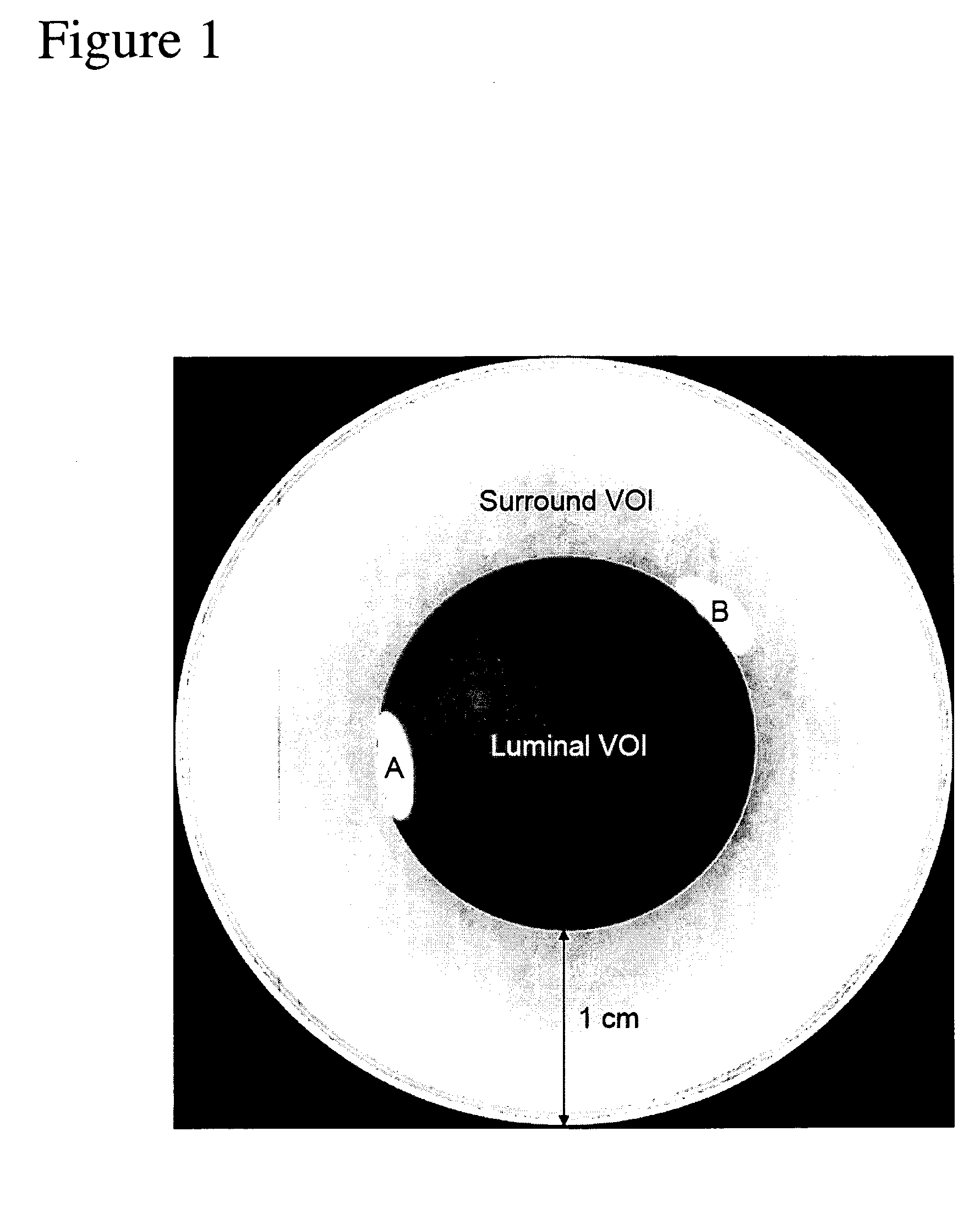
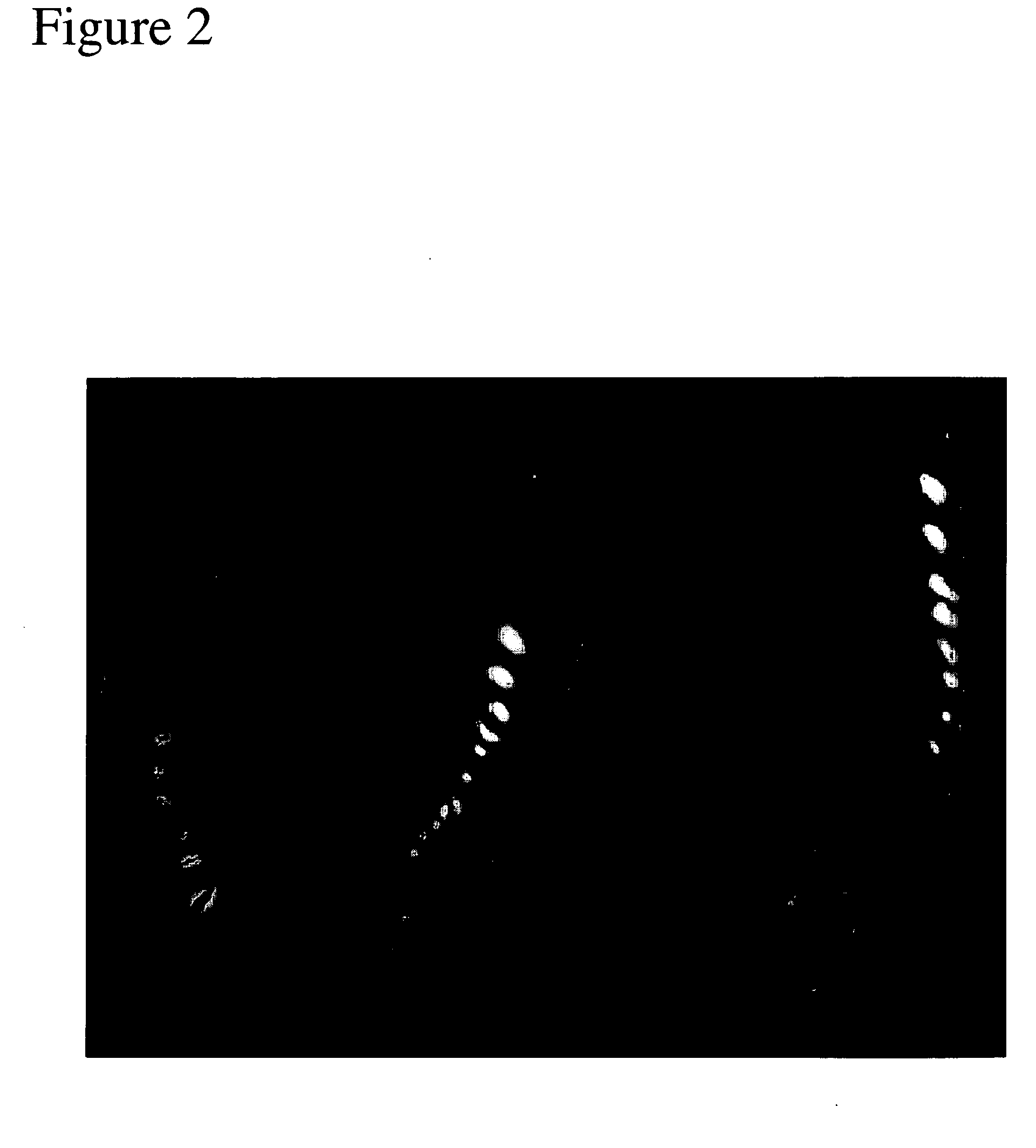
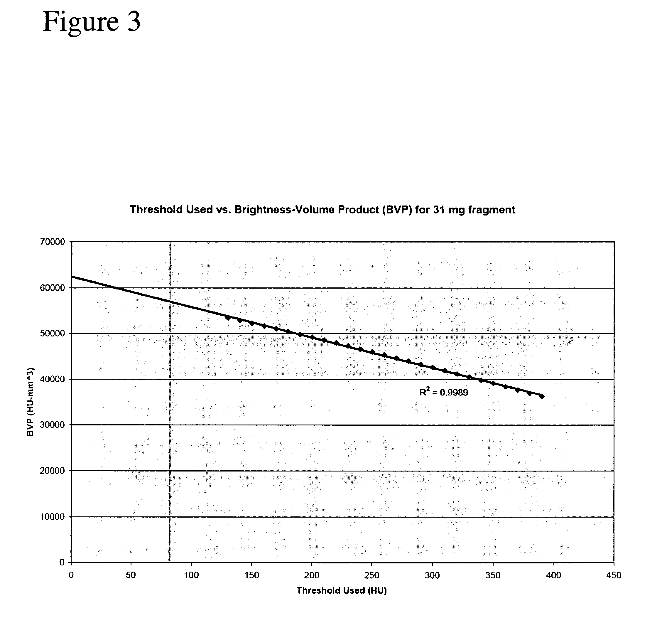
[0009] The present invention provides a method that allows the quantification of the true mass of a calcium fragment located inside or outside a vessel as well as with respect to a systemic artery. The method is independent of the level of arterial contrast enhancement, does not require protocol-specific or scanner-specific calibration scans, and allows a detailed analysis of calcium distribution patterns. To achieve these advantages, the method first identifies one or more calcium fragments in an image. In a preferred embodiment, the images are obtained Computed Tomography Angiography. For each of the identified calcium fragments the average intensity and volume is determined as a function of a plurality of intensity thresholds. Using these determined values, brightness volume products are calculated, which are defined as the product of the average intensity and the volume. These products are determined for each of the plurality of intensity thresholds. The mass of a calcium segment is subsequently obtained from the calculated brightness volume products extrapolated at zero intensity and reference calcium parameters. In an additional embodiment, the mass and volume of one or more calcium fragments are visualized with respect to a vessel in a computer display.

Problems solved by technology

The occurrence of iliac calcification was also associated with an increased incidence of symptoms of intermittent claudication.
However, these methods are threshold and protocol-specific, and may require calibration scans to obtain comparable measurements across different scanners and protocols and to correct for varying levels of arterial contrast enhancement (See e.g. Hong et al.
While these methods allow total mass quantification per vessel and per scan, they do not include a detailed analysis of the shape, size and distribution patterns of calcium fragments.
This presents an additional problem, as enhancement reduces the apparent volume of calcium fragments, potentially reducing the accuracy of mass- quantification.
Calcium quantification in the systemic arteries, as opposed to coronary arteries, also presents a unique challenge of practicality.
CT studies currently performed for quantifying calcification of the coronary arteries include less than 100 slices and take 10-15 minutes to process manually, CTAs currently consist of hundreds to thousands of images, and therefore take significantly longer.

Method used


Image

  • Quantification method of vessel calcification
  • Quantification method of vessel calcification
  • Quantification method of vessel calcification

Examples


Embodiment Construction

[0016] The present method requires either two-dimensional images or three-dimensional volume data sets acquired via computed tomographic angiography, MR imaging, or the like. The image(s) include(s) one or more vessels of interest that one would like to identify. Identification could for instance be accomplished by identifying the start and end-points of the vessel(s) of interest. In a preferred embodiment for vessel identification the bony structures are excluded from the image. A previously developed method by the same group as the present invention is suitable (i.e. U.S. patent application Ser. No. 10 / 723,166 with filing date of Nov. 26, 2003 which is hereby incorporated by reference for all that it discloses). This method is preferred and capable of removing structures that possess the shape and intensity distribution characteristic of bone. The resulting image is left with vessel structures from which median centerlines can be obtained (See Paik et al. 1998 in a paper entitled ...


PUM


Abstract

A method that allows the quantification of the true mass of a calcium fragment located along a vessel is provided. The method is independent of the level of arterial contrast enhancement, does not require protocol-specific or scanner-specific calibration scans, and allows a detailed analysis of calcium distribution patterns. For each identified calcium fragment, the average intensity and volume is determined as a function of a plurality of intensity thresholds. Using these determined values brightness volume products are calculated for each of the plurality of intensity thresholds. The mass of a calcium segment is subsequently obtained from the calculated brightness volume products extrapolated at zero intensity and reference calcium parameters. The mass and volume of the calcium fragments could be visualized with respect to a vessel in a computer display.

Description

CROSS-REFERENCE TO RELATED APPLICATIONS [0001] This application is cross-referenced to and claims priority from U.S. Provisional Application 60 / 526,790 filed Dec. 3, 2003, which is hereby incorporated by reference. STATEMENT REGARDING FEDERALLY SPONSORED RESEARCH OR DEVELOPMENT [0002] The present invention was supported in part by grant numbers 5R01HL58915 and 1R01HL67194 both from the National Institutes of Health (NIH). The U.S. Government has certain rights in the invention.FIELD OF THE INVENTION [0003] The present invention relates generally to medical imaging. More particularly, the present invention relates to methods of quantifying vascular calcification. BACKGROUND [0004] Vascular calcifications have been associated with the development of regional vascular disease. The severity of aortoiliac calcification as seen on chest x-ray studies has been correlated with atherosclerotic risk factors, coronary artery disease, peripheral arterial disease and cardiovascular mortality. Ao...

Claims


Application Information

Patent Timeline
IPC IPC(8): G21K1/12H05G1/60
CPCG06T7/0012G06T2207/30101G06T2207/10081G06T2200/24
Inventor RAMAN, RAGHAVRAMAN, BHARGAVNAPEL, SANDY A.RUBIN, GEOFFREY D.
Owner THE BOARD OF TRUSTEES OF THE LELAND STANFORD JUNIOR UNIV
Learn More