

Materials and method for promotion of nerve regeneration
a nerve regeneration and material technology, applied in the field of materials and methods for promoting nerve regeneration, can solve the problems of pain, neuronal death, muscle atrophy and permanent functional deficit, and little axon regeneration, so as to improve the ability of regenerating axons and potentiate axonal growth
- Summary
- Abstract
- Description
- Claims
- Application Information
AI Technical Summary
Benefits of technology
Problems solved by technology
Method used
Image

Examples
example 1
Composition of Marked Axons in thy-1-YFP-H Mice
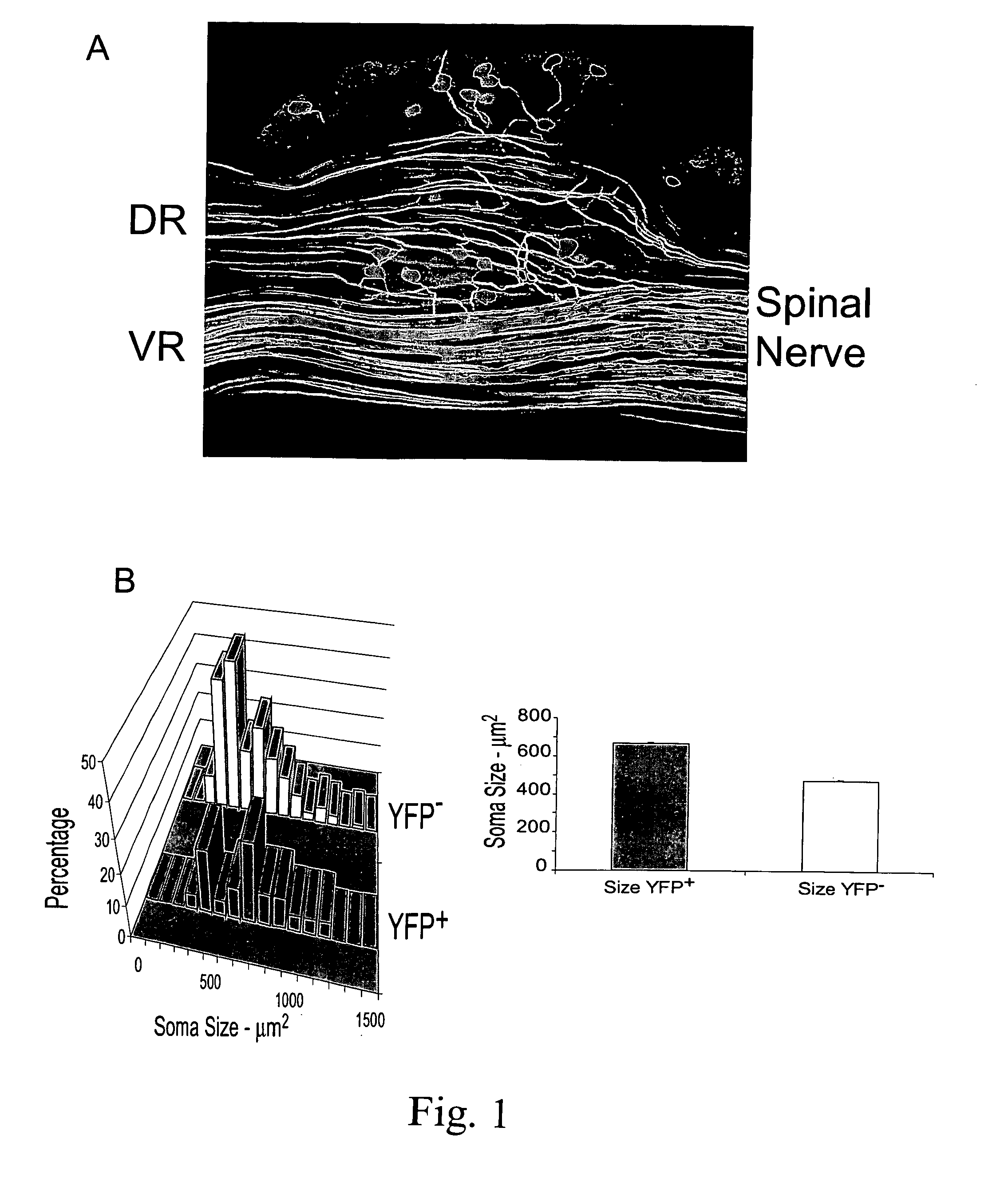
[0095] In the H strain of thy-1-YFP mice, a subset of axons in peripheral nerves is marked by the presence of yellow fluorescent protein (Feng G, Mellor R H, Bernstein M, Keller-Peck C, Nguyen Q T, Wallace M, Nerbonne J M, Lichtman J W, Sanes J R [2000]“Imaging neuronal subsets in transgenic mice expressing multiple spectral variants of GFP”Neuron 28:41-51), but the proportion of all axons marked, whether sensory or motor, is not known. The number of YFP+axons were counted in the dorsal and ventral roots of the main segmental nerves (L4 and L5) which contribute to the CF nerve in optical sections (FIG. 1A) through whole mounts of spinal nerves from three mice (five sets of nerves). The mean numbers of sensory and motor axons (±SEM) are shown in Table I.
TABLE 1Summary of YFP+ axons in spinal roots of thy-l-YFP-H miceAll values are means ± SEMDorsal Root AxonsVentral Root AxonsTotalL4122.60 ± 15.29(58%)87.20 ± 16.23(42%)209.80 ± 31.49L...
example 2
Axon Regeneration in the thy-1-YFP-H Mouse
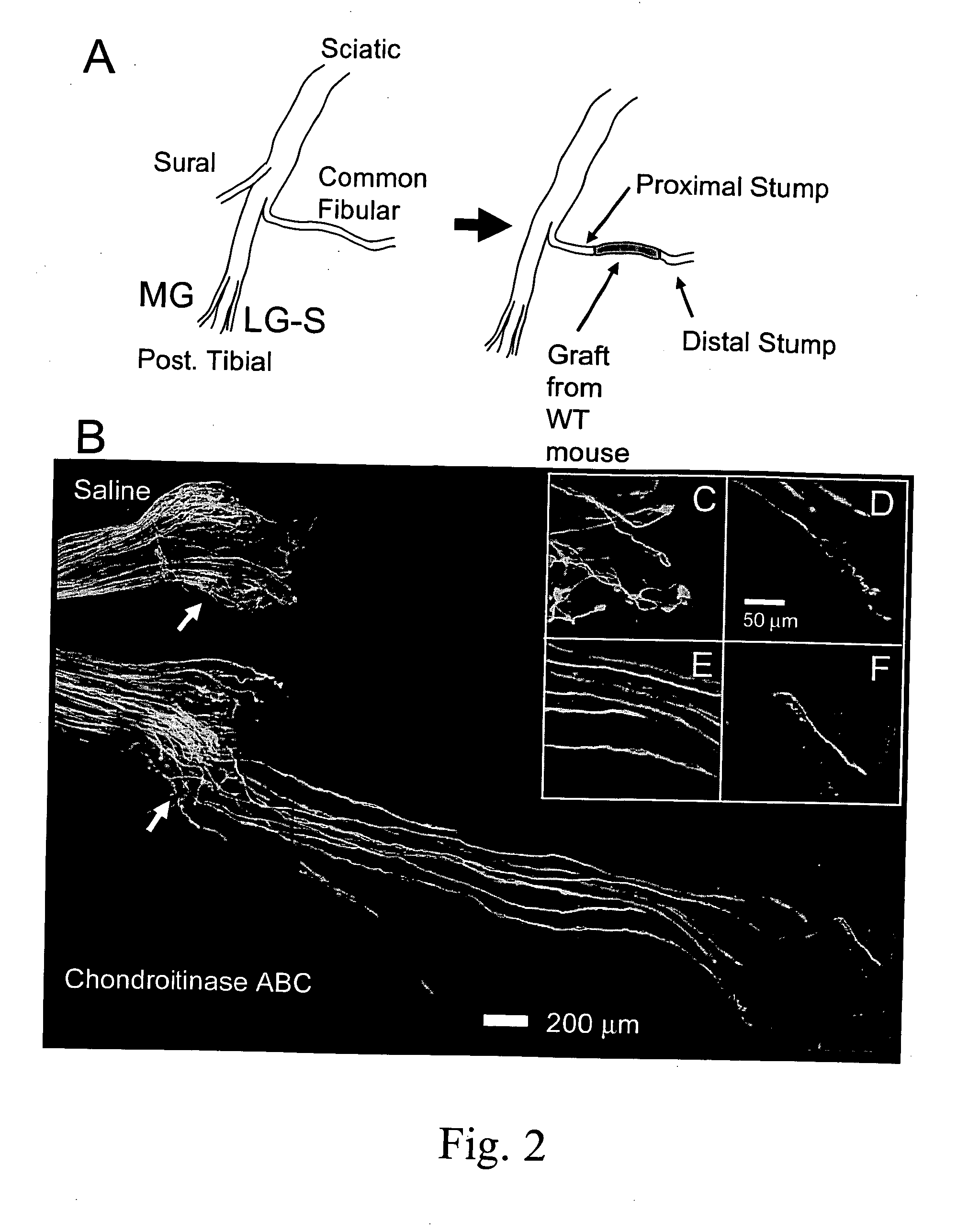
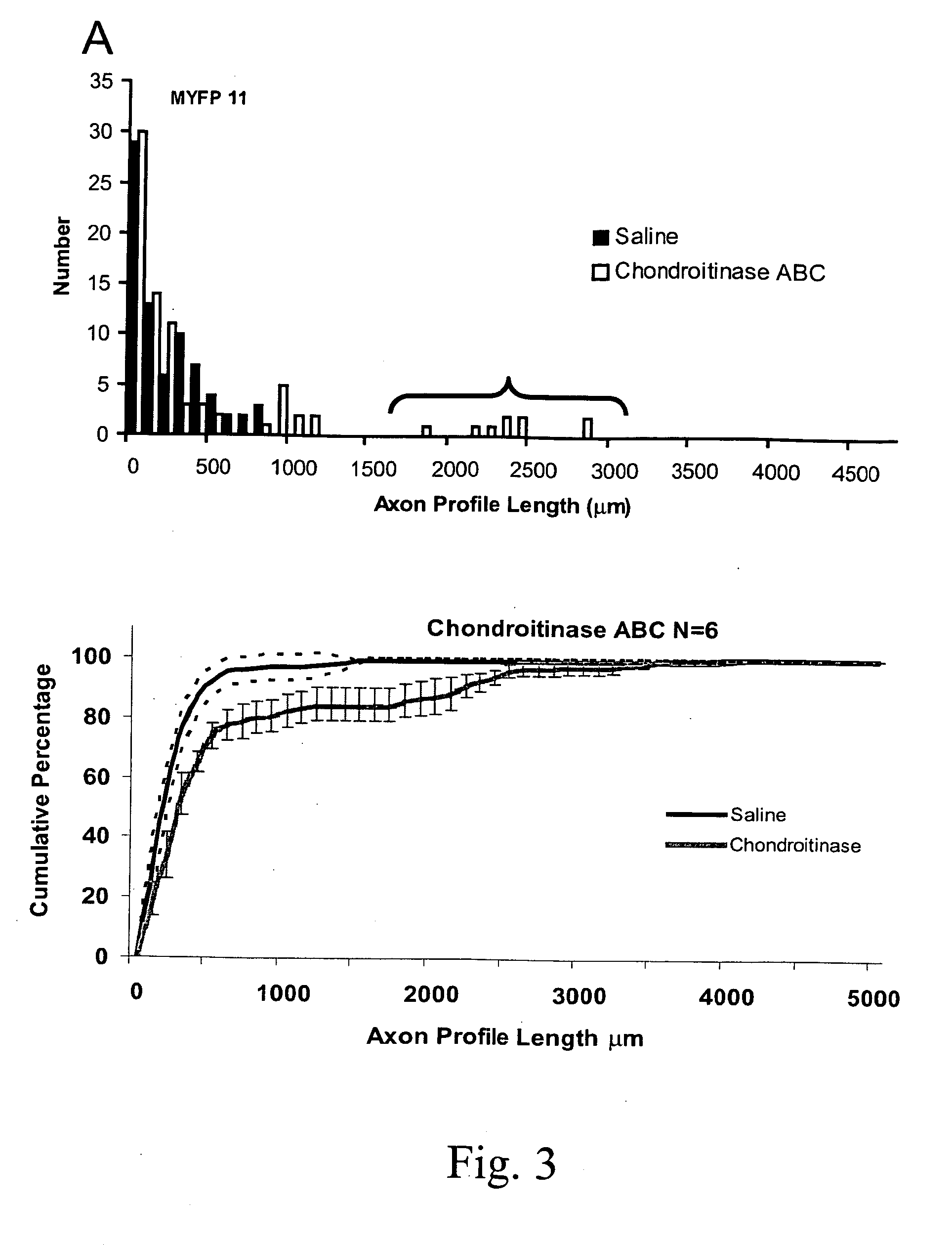
[0098] In thy-1-YFP-H mice one week after repairing a surgical transection of the CF nerve with an untreated or saline-treated graft from a wild type littermate (FIG. 2A), little evidence for axon regeneration is noted (FIG. 2B: Saline). One week after nerve repair, most of the fluorescent profiles of the marked axons are found close to the surgical repair site, the interface between the proximal stump of the cut nerve and the graft. A few axons have penetrated the graft and have grown into it, but most axon profiles observed have not. This lack of growth of axons into untreated grafts is reflected in the distribution of the measured lengths of axon profiles (FIG. 3A). Greater than 90% of fluorescent axon profiles in these nerves were shorter than 500 μm.
example 3
Chondroitinase ABC Treatment Enhances Axonal Regeneration
[0099] In thy-1-YFP-H mice one week after repairing the cut CF nerve with a graft from a wild type mouse which had been treated with chondroitinase ABC, the outcome is quite different (FIG. 2B, Chondroitinase ABC). Whereas, most of the axon profiles in saline-treated or untreated grafts ended in large flat endings which resemble either lamellopodia-containing growth cones or retraction bulbs (FIG. 2C), the endings of axon profiles that had entered and grown into the chondroitinase-treated grafts were more fusiform (FIG. 2D-F). Many more fluorescent axons are found to have grown into the graft, some for considerable distances. This observation is reflected in the distribution of the lengths of profiles of fluorescent regenerating axons. As shown in FIG. 3A, which is a histogram of measurements of axon profile lengths from the left and right nerves of a single mouse, many axon profile lengths in both saline-treated and chondroi...
PUM
| Property | Measurement | Unit |
|---|---|---|
| Fraction | aaaaa | aaaaa |
| Fraction | aaaaa | aaaaa |
| Fraction | aaaaa | aaaaa |
Abstract
Description
Claims
Application Information