Positive contrast MRI of magnetically tagged cells, objects, tissues

a magnetically tagged cell and positive contrast technology, applied in the field of magnetic resonance imaging, can solve the problems of significant signal dephasing, negative contrast agents suffering from partial volume effects, and agents cannot be distinguished from voids in images, and achieve the effect of positive contras

Active Publication Date: 2005-11-24
THE BOARD OF TRUSTEES OF THE LELAND STANFORD JUNIOR UNIV
View PDF5 Cites 22 Cited by
  • Summary
  • Abstract
  • Description
  • Claims
  • Application Information

AI Technical Summary

Benefits of technology

[0004] The present invention is a new method for imaging objects and materials that cause a localized magnetic field inhomogeneity in an MRI scanner with positive contrast. In one embodiment of the new method, spectrally-selective RF pulses are used to excite and refocus the off-resonance water surrounding the cells labeled wi

Problems solved by technology

Cells loaded with SPIO cause significant signal dephasing due to the magnetic field inhomogeneity induced in water molecules near the cell.
A fundamental drawback of negative contrast agents is that the agent cannot be distinguished from a void in the image.
Moreover, negative contrast agents suffer from partial-volume effects, where the ability to dete

Method used


Image

  • Positive contrast MRI of magnetically tagged cells, objects, tissues
  • Positive contrast MRI of magnetically tagged cells, objects, tissues
  • Positive contrast MRI of magnetically tagged cells, objects, tissues

Examples


Embodiment Construction

Theory

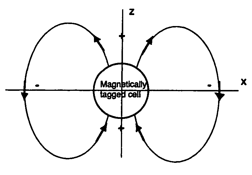
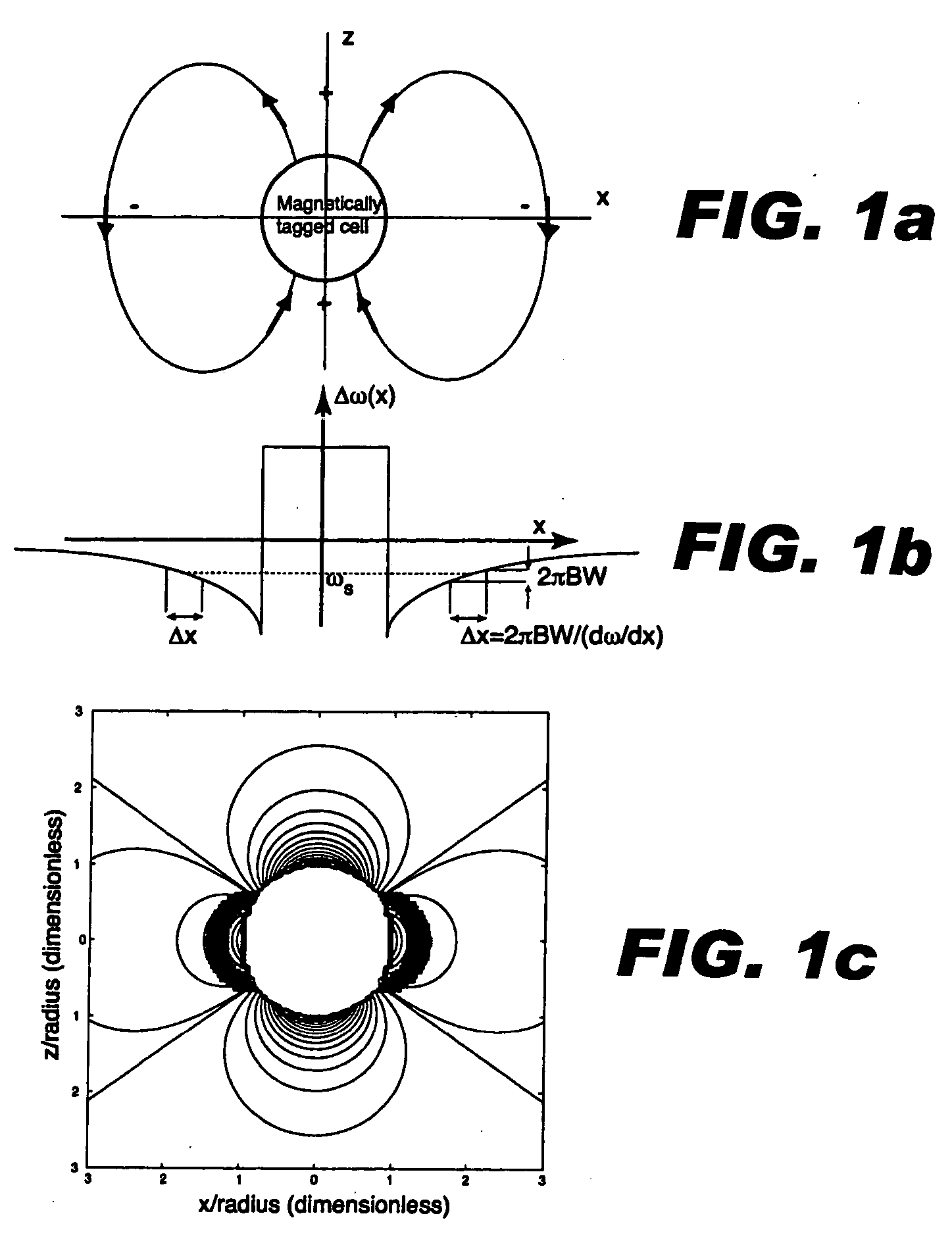
[0012] A collection of labeled cells will cast a field pattern in the water molecules immediately surrounding the cells. The field pattern can be approximated by a dipole field from a magnetized sphere. The dipole pattern demonstrates a classic field cross pattern, in which the local Bz field is enhanced in the north and south poles and suppressed along the equator. The polarity of the field perturbation would be reversed for a diamagnetic agent. The dipole field pattern intensity falls off quickly. The field perturbation varies as Δ⁢ ⁢Bz⁡(r,θ)=Δχ⁢ ⁢Bo3⁢(ar)3⁢(3⁢cos2⁢θ-1)(1)

where Δχ is the difference in bulk magnetic susceptibility between the sphere and surroundings, α is the radius of the sphere, r is the distance from the sphere center, and θ is the angle relative to the main field, Bo. Hence, the field pattern from a smaller collection of cells will fall off more steeply than that from a larger collection. In practice, aglomerations of labeled cells may not be spherica...


PUM


Abstract

Contrast agents incorporating super-paramagnetic iron-oxide (SPIO) nanoparticles have shown promise as a means to visualize labeled cells using MRI. Labeled cells cause significant signal dephasing due to the magnetic field inhomogeneity induced in water molecules near the cell. With the resulting signal void as the means for detection, the particles are behaving as a negative contrast agent, which can suffer from partial-volume effects. Disclosed is a new method for imaging labeled cells with positive contrast. Spectrally-selective RF pulses are used to excite and refocus the off-resonance water surrounding the labeled cells so that only the fluid and tissue immediately adjacent to the labeled cells are visible in the image. Phantom, in vitro, and in vivo experiments show the feasibility of the new method. A significant linear correlation (r=0.87, p<0.005) between the estimated number of cells and the signal has been observed.

Description

BACKGROUND OF THE INVENTION [0001] This invention relates generally to magnetic resonance imaging (MRI), and more particularly the invention relates to the use of MRI in tracking magnetically labeled cells or objects using positive contrast. INTRODUCTION [0002] Contrast agents incorporating super-paramagnetic iron-oxide (SPIO) nanoparticles have shown much promise as a means to visualize labeled cells using MRI. The small size of the particles (<100 nm) facilitates transport across cell membranes, and the low toxicity allows for large iron loads without significant cell death (e.g., 25 pg / cell). Labeling can be performed by incubating cells of interest (e.g., embryonic stem cells) with the contrast agent in vitro, so that they can be monitored in vivo using MRI. Cells such as macrophages can be labeled in vivo by introducing the contrast agent into the bloodstream, with the uptake of the agent occurring by phagocytosis, which has been used to image atherosclerosis and other infla...

Claims


Application Information

Patent Timeline
IPC IPC(8): A61B5/05A61B5/055A61B5/06
CPCA61B5/055G01R33/5601A61B5/06
Inventor CONOLLY, STEVEN M.CUNNINGHAM, CHARLES H.
Owner THE BOARD OF TRUSTEES OF THE LELAND STANFORD JUNIOR UNIV
Why Eureka
Try Eureka