
Magnetic resonance method and apparatus for acquisition of image data of a vessel wall

a magnetic resonance and image data technology, applied in the field of magnetic resonance method and apparatus for acquisition of image data of vessel walls, can solve the problems of insufficient detection of calcifications and/or calcium deposits in plaques, insufficient evaluation of at-risk patients, and high cost, so as to improve the evaluation of composition and improve the identification of patients.

Inactive Publication Date: 2008-06-26
SIEMENS AG
View PDF3 Cites 17 Cited by
  • Summary
  • Abstract
  • Description
  • Claims
  • Application Information

AI Technical Summary

Benefits of technology

[0014]An object of the present invention is to provide a method for acquisition of an image of a vessel wall that enables a non-invasive, x-ray-free and high-resolution image acquisition with which an image of an atherosclerotic vessel wall variation can be acquired. The method should allow an improved evaluation of the composition of the vessel wall variation and an improved identification of patients at risk for a thromboembolic event. Furthermore, it is an object of the invention to provide a magnetic resonance apparatus for implementation of such a method.
[0019]It is possible to also measure signals of tissue components with a short T2 relaxation time (such as, for example, calcified tissue) so that this tissue also generates a positive contrast (i.e. a visible signal) in the image. In the generated image this is advantageous since now calcifications (which generate only a negative contrast with conventional MR sequences, i.e. generate only an insufficient signal in the representation) can be made visible. The generated image allows a user to better assess the composition of a vessel wall variation. Computer-aided evaluation algorithms based on the generated image data can likewise implement a more precise quantification of tissue components of a plaque since now one of the components that is essential for a diagnosis of the vulnerability of a plaque, namely calcifications or calcium deposits, generates a distinctly visible and measurable signal.
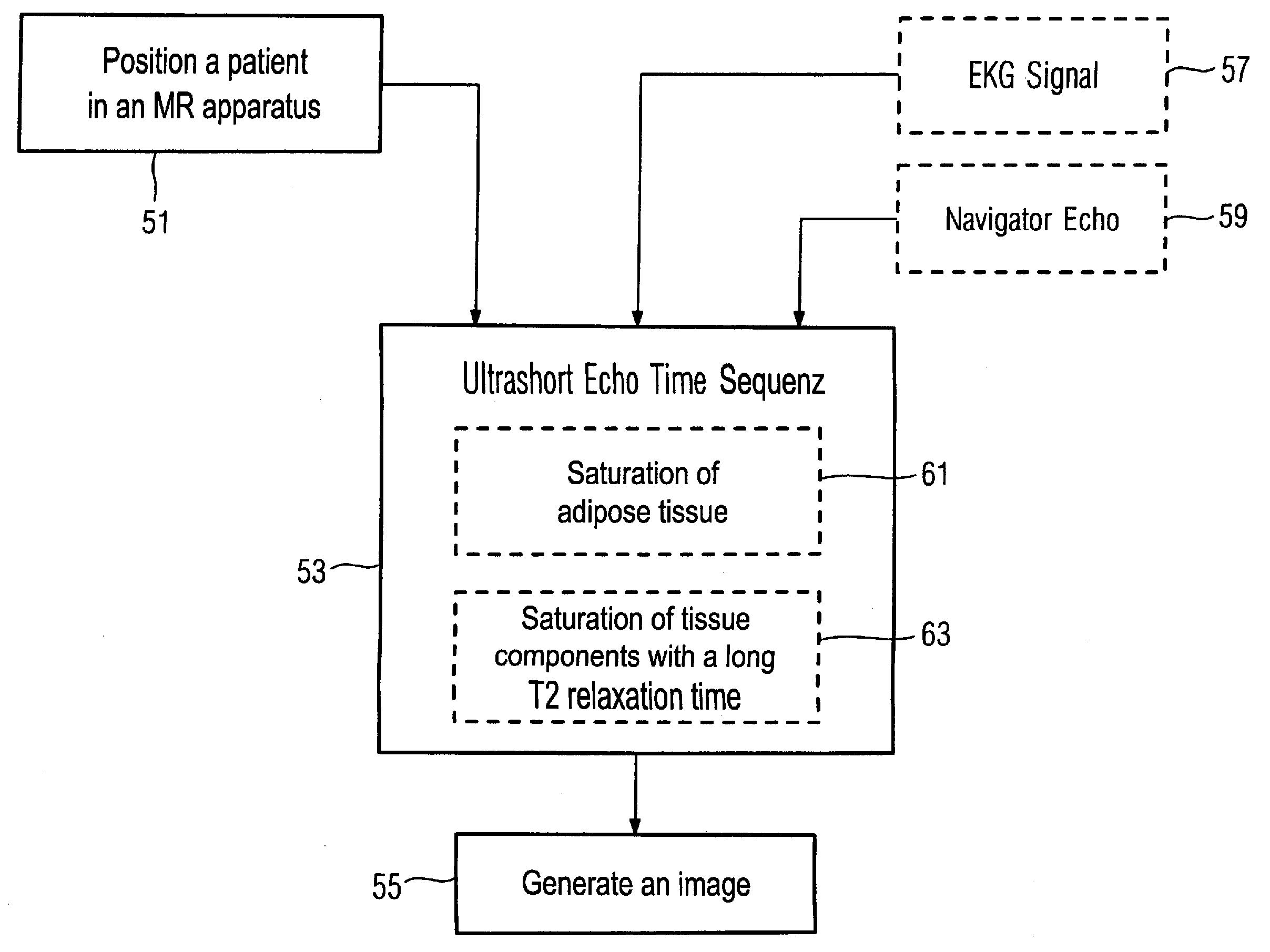
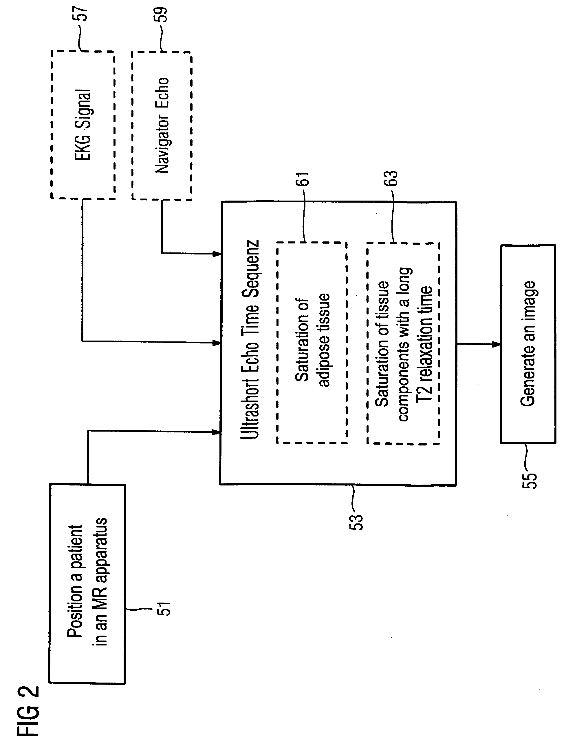
[0020]In an embodiment, the ultrashort echo time sequence includes at least one radio-frequency saturation pulse for suppression of signals of nuclear spins of fat tissue. In this embodiment, it is possible to reduce signals that have their origin in nuclear spins of fat tissue since these nuclear spins are saturated by the radio-frequency saturation pulse. The contrast between lipid deposits and calcifications in a vessel wall hereby increases.
[0021]In another embodiment, the ultrashort echo time sequence includes at least one radio-frequency saturation pulse for suppression of signals of nuclear spins whose T2 relaxation time is greater than a predetermined threshold. It is thereby possible to reduce signals that have their origin in tissue with a long T2 relaxation time. In the generated image this causes a higher contrast between this type of tissue and calcified tissue.
[0022]K-space is advantageously three-dimensionally scanned with the ultrashort echo time sequence. The scanning of k-space preferably ensues in a radial manner. Such a scanning trajectory shows a relatively low susceptibility to movement artifacts and additionally allows the production of an image with a small image region FOV (field of view) with high resolution.

Problems solved by technology

Intravascular ultrasound (IVUS) allows a radiation-free examination of the vessel wall, and is predominantly suitable for soft, non-calcified plaque, but is an invasive examination method and is relatively expensive.
The size of the lumen, however, does not correlate with the vulnerability of a plaque to rupture, which is why at-risk patients can be only insufficiently identified with this examination method.
Further analysis of this approach has shown that calcifications and / or calcium deposits in a plaque can be only insufficiently detected since calcium, due to its short T2 relaxation time, appears in the image as a region with signal attenuation.
Signal attenuations, however, can also be based on various artifacts, such that calcifications are often overestimated.

Method used


Image

  • Magnetic resonance method and apparatus for acquisition of image data of a vessel wall
  • Magnetic resonance method and apparatus for acquisition of image data of a vessel wall
  • Magnetic resonance method and apparatus for acquisition of image data of a vessel wall

Examples


Embodiment Construction

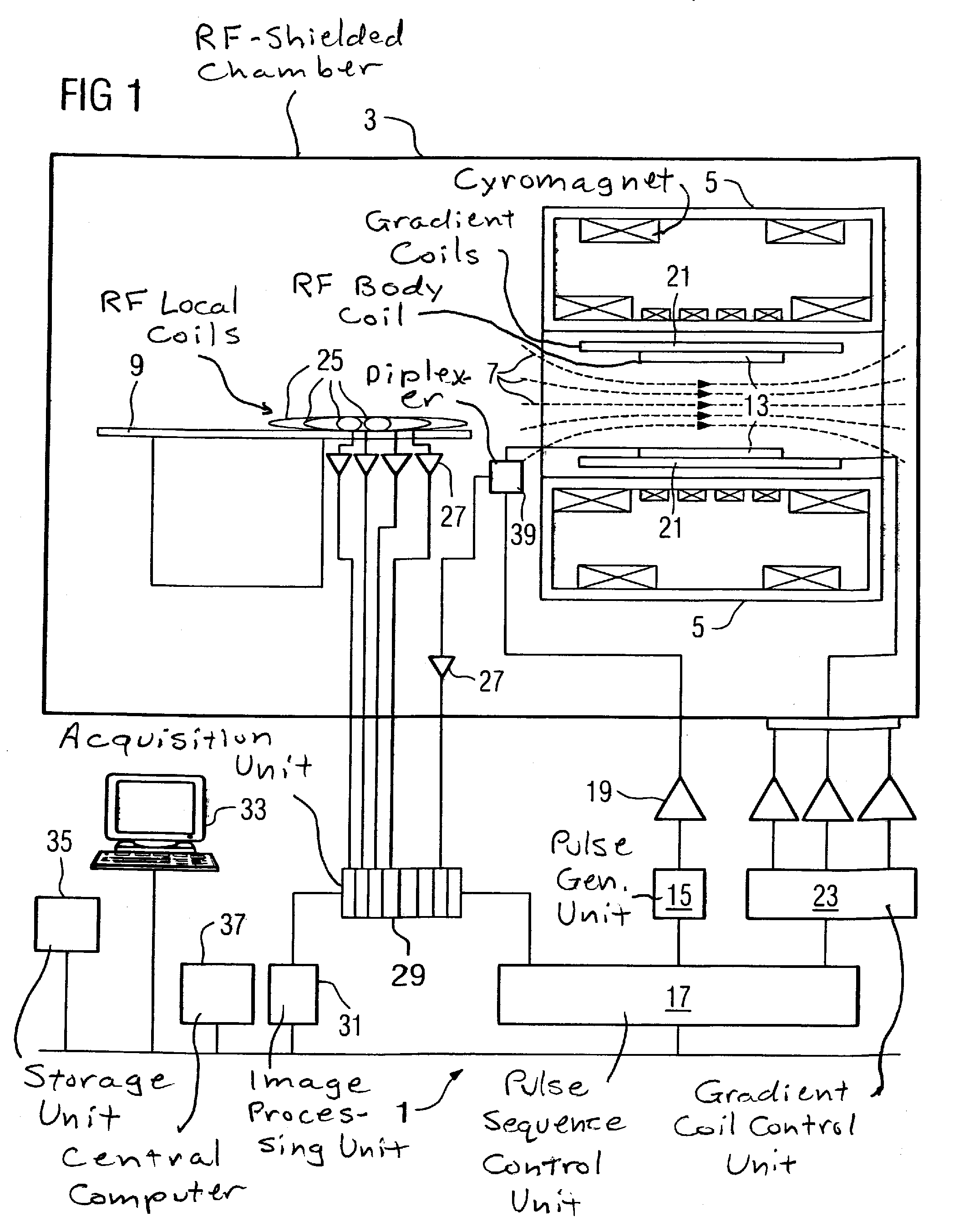
[0030]FIG. 1 schematically shows the basic design of a magnetic resonance apparatus 1. In order to examine a body by means of magnetic resonance imaging, various magnetic fields matched as precisely as possible to one another in terms of their temporal and spatial characteristics are applied.

[0031]A strong magnet (typically a cryomagnet 5 with a tunnel-shaped opening) arranged in a radio-frequency-shielded measurement chamber 3 generates a static, strong basic magnetic field 7 that is typically 0.2 Tesla to 3 Tesla and more. A body or a body part (not shown) to be examined is surprised on a patient bed 9 and positioned in a homogeneous region of the basic magnetic field 7.

[0032]The excitation of the nuclear spins of the body ensues by radio-frequency excitation pulses that are radiated by a radio-frequency antenna (shown here as a body coil 13). The radio-frequency excitation pulses are generated by a pulse generation unit 15 that is controlled by a pulse sequence control unit 17. A...


PUM


Abstract

In a magnetic resonance method and apparatus for acquisition of an image for examination of a vessel wall variation, a vessel wall section of a patient to be examined is positioned in an imaging volume of the magnetic resonance apparatus, image data of the vessel wall section are acquired with an ultrashort echo time sequence, and an image is generated from the acquired image data.

Description

BACKGROUND OF THE INVENTION[0001]1. Field of the Invention[0002]The present invention concerns a method for acquisition of image data of a vessel wall by means of magnetic resonance technology as is used for diagnosis of variations of the vessel wall that are due to an atherosclerosis. The invention also concerns a magnetic resonance apparatus for implementing such a method.[0003]2. Description of the Prior Art[0004]Arteriosclerosis is a systemic illness of the arteries that leads to deposits of blood lipids, thromboses, connective tissue and calcium in the vessel walls. The focal variations that occur in the inner and in the middle vessel wall are also referred to as atherosclerosis. The atherosclerotic variations are often locally limited and form what are known as plaques. Among other things, heart infarcts and strokes are among the typical results of arteriosclerosis.[0005]Thromboembolic events, i.e. the formation of a blood clot in an artery, are often based on a rupture of a “...

Claims


Application Information

Patent Timeline
Patent Type & Authority Applications(United States)
IPC IPC(8): A61B5/055
CPCA61B5/02007A61B5/055A61B5/721A61B5/7292G01N24/081G01R33/4816G01R33/5676G01R33/5602G01R33/5607G01R33/5615G01R33/5616G01R33/5673G01R33/4824
Inventor NIELLES-VALLESPIN, SONIA
Owner SIEMENS AG
Learn More