Method and system for digital image based flourescent in situ hybridization (FISH) analysis

a technology of flourescent in situ hybridization and digital image, applied in the field of digital image processing, can solve the problems of not giving enough information for the referring medical clinician to make decisions about patient prognosis and treatment, the difficulty of simple task of counting cells of interest, and the possibility of missing genuine cells of interest by experienced pathologists

Inactive Publication Date: 2005-12-01
BIOIMAGENE
View PDF2 Cites 62 Cited by
  • Summary
  • Abstract
  • Description
  • Claims
  • Application Information

AI Technical Summary

Problems solved by technology

In many areas of histopathology, such as a diagnosis of breast carcinoma, does not give enough information for the referring medical clinician to make decisions about patient prognosis and treatment.
It is observed that the seemingly simple task of counting cells of interest becomes difficult because the counting has to be done for large number of sections.
Even experienced pathologist might miss genuine cells of interest due to fatigue.
However, many images do not have pixel values that make effective use of the full dynamic range of pixel values available on an output device.
The result in either case is that the output is relatively dull in appearance.
Clinical studies in patients with breast cancer over the last decade have convincingly demonstrated that amplification/ overexpression of Her-2/neu is associated with a poor prognosis.
However, in practice, touching, overlapping nuclei is a major technical problem that image processing algorithms should address.
Reported accuracy of 90% is not sufficient for field leve

Method used


Image

  • Method and system for digital image based flourescent in situ hybridization (FISH) analysis
  • Method and system for digital image based flourescent in situ hybridization (FISH) analysis
  • Method and system for digital image based flourescent in situ hybridization (FISH) analysis

Examples


Embodiment Construction

Exemplary Fluorescence In Situ Hybridization (FISH) Analysis System

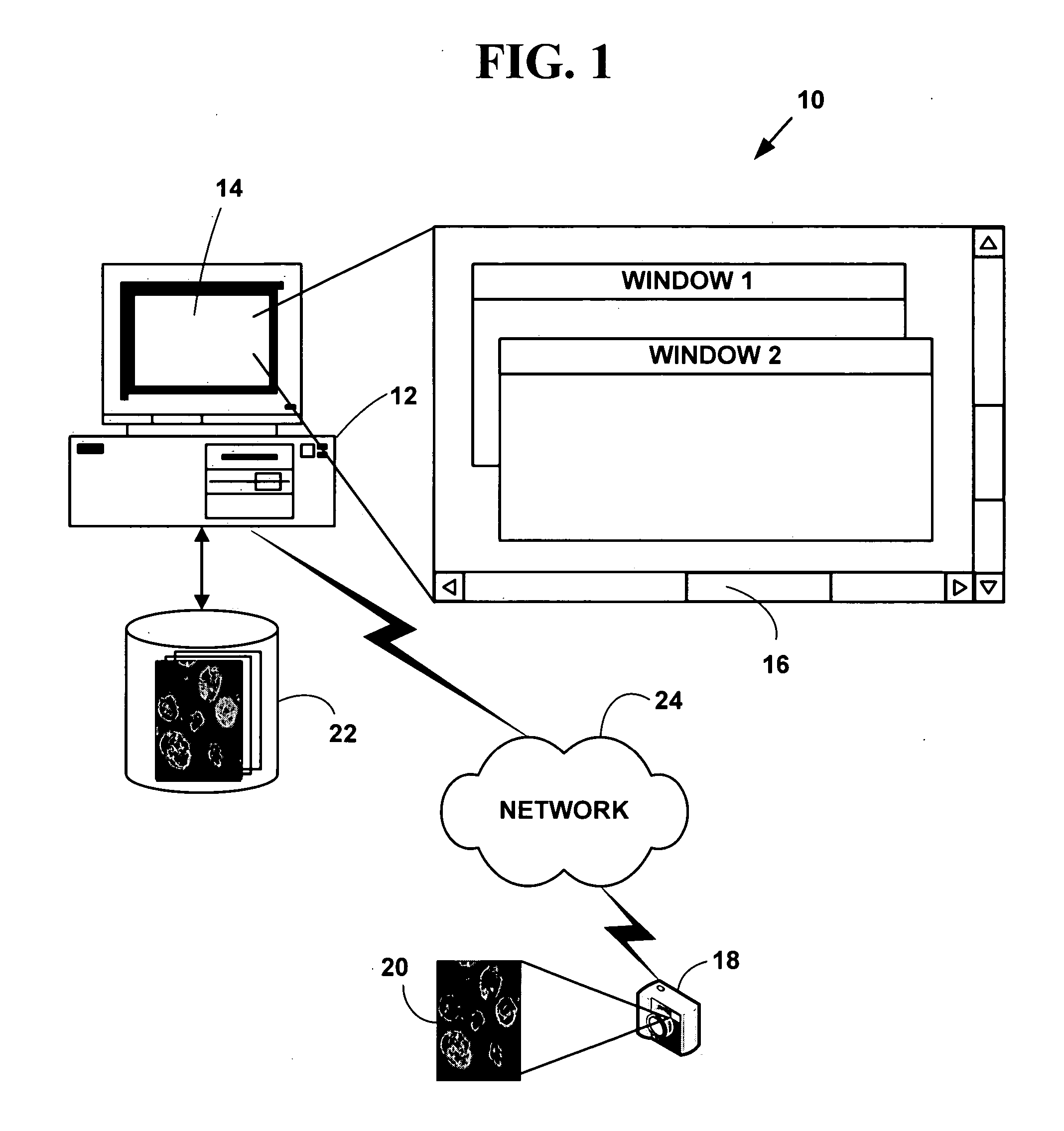
[0050]FIG. 1 is a block diagram illustrating an exemplary automated fluorescence in situ hybridization (FISH) analysis system 10. The exemplary system 10 includes one or more computers 12 with a display 14 (one of which is illustrated). The display 14 presents a windowed graphical user interface (“GUI”) 16 with multiple windows to a user. The system 10 may optionally include an optical or fluorescent microscope or other magnifying device (not illustrated in FIG. 1).

[0051] As is known in the art, a conventional “optical microscope” uses light to illuminate a sample and produces a magnified image of the sample. A “fluorescence microscope” uses a much higher intensity light to illuminate the sample. This light excites fluorescent compounds in the sample, which then emit light of a longer wavelength. A fluorescent microscope also produces a magnified image of the sample, but the image is based on the second light sourc...


PUM


Abstract

A method and system for automated digital fluorescent in situ hybridization (FISH) image analysis. Luminance parameters from a digital image of a biological tissue sample to which a fluorescent compound (e.g., LSI-HER-2/neu and CEP-17 dyes) have been applied are analyzed to determine plural regions of interest. Fluorescent color signals in the plural regions of interest including plural cell nuclei are identified, classified and grouped into plural groups. Each of the plural groups is validated based on pre-defined conditions. A medical diagnosis or prognosis or medical, life science or biotechnology experiment conclusion determined using a count of plural ratios of validated fluorescent color signals within each of the cell nuclei within the plural groups.

Description

CROSS REFERENCES TO RELATED APPLICATIONS [0001] This application claims priority to U.S. Provisional Application to U.S. Provisional patent application No. 60 / 541,301, filed Feb. 3, 2004. This application also claims priority to U.S. patent application Ser. No. 10 / 938,314, filed Sep. 10, 2004, which claims priority U.S. Provisional Patent Application No. 60 / 501,142, filed Sep. 10, 2003, and U.S. Provisional Patent Application No. 60 / 515,582 filed Oct. 30, 2003, and U.S. patent application Ser. No. 10 / 966,071, filed Oct. 23, 2004 which claims priority to U.S. Provisional Patent Application No. 60 / 530,714, filed Dec. 18, 2003, the contents of all of which are incorporated by reference.COPYRIGHT NOTICE [0002] Pursuant to 37 C.F.R. 1.71(e), applicants note that a portion of this disclosure contains material that is subject to and for which is claimed copyright protection, such as, but not limited to, digital photographs, screen shots, user interfaces, or any other aspects of this submis...

Claims


Application Information

Patent Timeline
IPC IPC(8): G06K9/00G06K9/46
CPCG06K9/00127G06V20/69
Inventor GHOLAP, ABHIJEET S.GHOLAP, GAURI A.RAO, C. V.K.VIPRA, MADHURAPARDESHI, DEEPAK M.JADHAV, PRITHVIRAJABHYANKAR, JAYANT
Owner BIOIMAGENE
Why Eureka
Try Eureka