Guided access to lung tissues

a lung tissue and bronchoscopy technology, applied in the field of bronchoscopy working channel guide, can solve the problems of limited instrument size, limited scope use, limited working channel, etc., and achieve the effect of facilitating the spread of anatomical features

Inactive Publication Date: 2005-12-29
EKOS CORP
View PDF99 Cites 273 Cited by
  • Summary
  • Abstract
  • Description
  • Claims
  • Application Information

AI Technical Summary

Benefits of technology

[0016] Accordingly, the present invention provides a method of fixating a lung tissue for treatment or diagnosis. This method involves (a) delivering a lung access assembly into an inner part of the lung or surrounding tissue, wherein said assembly comprises (i) an imaging device comprising a working channel; and (ii) a guide element observable under a view of said imaging device, wherein at least a portion of said guide element is disposed inside said working channel to effect a guided access to said inner part or surrounding tissue of the lung with a plurality of fixation instruments, and wherein at least one fixation instrument of said plurality is operatively connected to a guide element; and (b) contacting said plurality of fixation instruments to fixate said lung tissue. In one aspect of this embodiment, the fixation instruments comprise needle guides carried therein a plurality of needles. Preferred needles are covered by an expandable sleeve.
[0017] The present invention further provides a space-making device for accessing a bodily organ or tissue. The device comprises the following components: (a) an elongated access device having a distal end, a proximal end, and a lumen therethrough, said device carrying a delivery element extendable through said lumen; (b) an open-ended and extendable sleeve surrounding said distal end of said elongated access device, wherein said extendable sleeve is designed to effect expansion of working space for accessing said bodily organ or tissue with said delivery element.
[0018] Also included in the present invention is a method of providing working space for accessing said bodily organ or tissue in a subject. The method involves (a) positioning a space-making device into said bodily organ or tissue of said subject, wherein said device comprises (i) an elongated access device having a distal end, a proximal end, and a lumen therethrough, said device carrying a delivery element extendable through said lumen; and (ii) an open-ended and extendable sleeve surrounding said distal end of said elongated access device; and (b) expending said extendable sleeve to effect expansion of working space for accessing said bodily organ or tissue with said delivery element.
[0019] In some aspects of this embodiment, the sleeve is radially extendable. In other aspects, the sleeve comprises at least one wing structure to facilitate spreading apart anatomical features at said bodily organ or tissue.

Problems solved by technology

The size of the working channel of conventional bronchoscopes limits the size of instruments that may be passed down the working channel to view, biopsy or treat a patient's lung at the distal end of the bronchoscope.
In addition, because the bronchoscopes working channel is being used to deliver the biopsy needle, the scope cannot be simultaneously used for other purposes, such as fixation of the target tissue.

Method used


Image

  • Guided access to lung tissues
  • Guided access to lung tissues
  • Guided access to lung tissues

Examples


Embodiment Construction

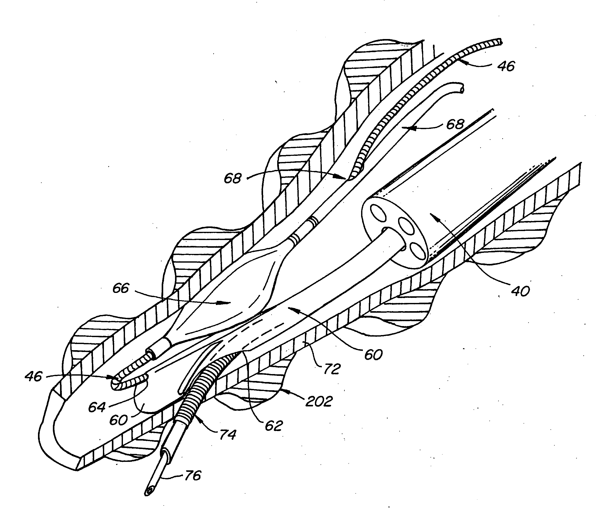
[0021]FIG. 5 shows a flexible bronchoscope 40 with a working channel 42 into which a needle guide 44 has been inserted. Prior to inserting bronchoscope 40 into a patient, a an access accessory such as guide wire 46 is inserted into the distal end 48 of needle guide 44. Guide wire 46 is bent around so that a proximal end 50 lies along the length of bronchoscope 40. When bronchoscope 40 is inserted into a patient's lungs, the proximal end 50 of guide wire 46 will remain outside of the patient. Guide wire 46 can then be used to deliver diagnostic, therapy or biopsy tools to the distal end of bronchoscope 40 without having to pass such tools through working channel 42. Such tools can be delivered either simultaneously alongside the bronchoscope or after the bronchoscope has been placed at the selected site within the patient's lung.

[0022] The guide wire 46 can also be used to position and steer the distal end 41 of the bronchoscope. Pulling guide wire 46 in a proximal direction will ca...


PUM


Abstract

This invention relates generally to lung access devices and methods of using the devices to gain access to the interior of a lung or to the mediastinal space around the lung. In particular, the invention relates to auxiliary access devices and tools for use with conventional bronchoscopes or other endoscopes to enable the delivery of more and larger devices to a target site than is currently possible through a typical endoscope or bronchoscope.

Description

CROSS-REFERENCE TO RELATED APPLICATIONS [0001] This application claims the priority benefit of U.S. Provisional Application Ser. No. 60 / 579,905 filed Jun. 14, 2004, which is hereby incorporated herein by reference in its entirety.BACKGROUND OF THE INVENTION [0002] This invention relates generally to the use of bronchoscopes and other devices to gain access to the interior of a lung or to the mediastinal space around the lung. In particular, the invention relates to auxiliary access devices and tools for use with conventional bronchoscopes or other endoscopes to enable the delivery of more and larger devices to a target site than is currently possible through a typical endoscope or bronchoscope. [0003] Most bronchoscopy cases use the bronchoscope simply as a tool to access the bronchi (transnasal, oral or trachea access to the lung) and possibly visualize abnormal colors from adjacent pathologic tissue. Most bronchoscope-based biopsies target tissues that lie outside the bronchi trun...

Claims


Application Information

Patent Timeline
Patent Type & Authority Applications(United States)
IPC IPC(8): A61B1/00A61B1/018A61B1/06A61B1/267A61B10/04A61B17/34
CPCA61B1/0014A61B1/018A61B2010/045A61B17/3403A61B1/2676
Inventor MATHIS, MARK
Owner EKOS CORP
Why Eureka
Try Eureka