Lung and lung cancer tissue culture method and method using lung and lung cancer tissue culture method to build lung cancer mouse animal model

A lung tissue and lung cancer technology, applied in the field of biomedicine, can solve the problems such as the inability of the PDX model to provide the in situ microenvironment of the lung tissue, the loss of biological characteristics of human-derived tumors, and the inability to meet the needs of model construction, achieving a high degree of consistency and quantity. It is easy to expand and is beneficial to the effect of scientific experimental research needs

Active Publication Date: 2017-07-21
WEST CHINA HOSPITAL SICHUAN UNIV
View PDF6 Cites 59 Cited by
  • Summary
  • Abstract
  • Description
  • Claims
  • Application Information

AI Technical Summary

Problems solved by technology

[0010] Since the results of mouse tumor genome research are far from directly being used to guide human tumor genomics research; while cell line xenograft tumor models are limited to differences in gene and biological functions between cell lines and human tumor cells; PDX Although the model partially compensates for the defects of the above two models, and is currently the main method for studying the biological characteristics of human tumors or drug screening and identification, it still has limitations. Due to the subcutaneous transplantation, the traditional PDX model cannot provide The in situ microenviron

Method used


Image

  • Lung and lung cancer tissue culture method and method using lung and lung cancer tissue culture method to build lung cancer mouse animal model
  • Lung and lung cancer tissue culture method and method using lung and lung cancer tissue culture method to build lung cancer mouse animal model
  • Lung and lung cancer tissue culture method and method using lung and lung cancer tissue culture method to build lung cancer mouse animal model

Examples


Embodiment 1

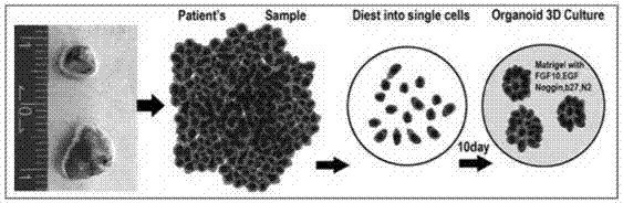
[0091] Lung Tissue and Lung Cancer Tissue Organoid Culture

[0092] as per figure 1 The process sequence shown is for 3D culture of lung tissue / lung cancer tissue organoids. Take lung tissue and lung cancer tissue and cut them into pieces on ice, add 10ml collagenase to resuspend, and run Human Lung and Human Tumor program 1 in gentleMACS C tube collagenase. Transfer to 37°C, 220rpm shaker for digestion for 20min. The Human Lung and Human Tumor programs 2 were run using gentalMACS. Transfer to 37°C, 220rpm shaker for digestion for 10min, filter the cells with a 100μm cell mesh, add 10ml DMEM / F12 to the filtrate to stop digestion, centrifuge (4°C, 200g, 5min), and remove the supernatant.

[0093] Take 5ml of erythrocyte lysate to resuspend, and lyse the erythrocytes on ice for 5min. In a centrifuge at 4°C, centrifuge at 200g for 5min, and remove the supernatant. Add 10ml DMEM / F12 to resuspend, centrifuge at 200°C for 5min at 4°C, and remove the supernatant. Obtain indep...

Embodiment 2

[0104] Lung Tissue and Lung Cancer Tissue Organoid Culture

[0105] Take lung tissue and lung cancer tissue and cut them into pieces on ice, add 10ml collagenase to resuspend, and run Human Lung and Human Tumor program 1 in gentleMACS C tube collagenase. Transfer to 37°C, 220rpm shaker for digestion for 15min. The Human Lung and Human Tumor programs 2 were run using gentalMACS. Transfer to 37°C, 220rpm shaker for digestion for 10min, filter the cells with a 100μm cell mesh, add 10ml DMEM / F12 to the filtrate to stop digestion, centrifuge (4°C, 200g, 6min), and remove the supernatant.

[0106] Take 5ml of erythrocyte lysate to resuspend, and lyse the erythrocytes on ice for 6min. In a centrifuge at 4°C, centrifuge at 200g for 5min, and remove the supernatant. Add 10ml DMEM / F12 to resuspend, centrifuge at 200°C for 7min at 4°C, and remove the supernatant.

[0107] For cell counting, mix Martrigel, 20,000 cells per 40 μL, drop in the center of a 64-well plate, place the cult...

Embodiment 3

[0111] Organoid Passaging

[0112] Collect the organoids in the culture dish of Example 2, resuspend the organoids in 3ml TrypLE, and digest at 37°C for 10min. Add 5ml DMEM / F12 to stop the digestion, centrifuge at 200g, 4°C for 5min, add an appropriate amount of Martrigel to resuspend according to the amount of cells, and drop in the center of the well of a 48-well plate. Place the Petri dish at 37°C (5% CO 2 ) 10min, solidify the Martrigel. Then, 150 μL of conditioned medium was added to each well at 37°C, 5% CO 2 Cell culture incubator. Change the medium every 2-3 days. Obtain passaged human lung tissue and lung cancer tissue passaged organoids.


PUM


Abstract

The invention discloses a method for culturing normal human lung tissue and a lung cancer tissue organoid in an in-vitro manner. The method includes: acquiring fresh human-derived lung tissue cells, and performing collagen digestion on the fresh human-derived lung tissue cells to obtain single cells; culturing human lung tissue and the lung cancer tissue organoid under in-vitro 3D culture conditions; performing H&E staining to determining the structure and form, and using q-PCR to detect related gene expression; using immunofluorescent staining to authenticate cell sources and detect related protein expression. The invention further discloses a method for building a mouse animal model based on the organoid. The method for culturing the normal human lung tissue and the lung cancer tissue organoid and the method for building the mouse animal model have the advantages that the methods are significant to the building of large-scale and good-consistency human-derived in-situ lung cancer animal models, and a good basis and related application prospect are provided for the fundamental researches of lung cancer.

Description

technical field [0001] The invention belongs to the field of biomedicine, in particular to a human lung normal tissue and lung cancer tissue organoid culture system, and belongs to cell culture or tissue culture technology. The invention also includes a method for constructing a mouse animal model by using the cultured lung cancer tissue. Background technique [0002] Lung cancer is currently the malignant tumor with the highest morbidity and mortality rate. The primary culture of lung tissue / lung cancer tissue cells and animal models of lung cancer are important tools for studying the occurrence, development and treatment of lung cancer. [0003] The traditional two-dimensional cell culture technology makes the cultured cells gradually lose their original state in the body in vitro, and their morphology, biological function, genetics and other aspects are very different from those in the human body, which cannot meet the existing requirements for Human lung cancer medical ...

Claims


Application Information

Patent Timeline
IPC IPC(8): C12N5/071C12N5/09A61K35/13A01K67/027
CPCA01K67/0271A01K2227/105A01K2267/0331A61K35/13C12N5/0062C12N5/0688C12N5/0693C12N2509/00C12N2513/00
Inventor 陈崇纳飞飞
Owner WEST CHINA HOSPITAL SICHUAN UNIV
Why Eureka
Try Eureka