
Method, system, and computer software product for feature-based correlation of lesions from multiple images

Active Publication Date: 2006-01-05
UNIVERSITY OF CHICAGO
View PDF15 Cites 50 Cited by
  • Summary
  • Abstract
  • Description
  • Claims
  • Application Information

AI Technical Summary

Benefits of technology

[0050] Embodiments of the present invention provide a classification scheme for an automated matching of lesions from multiple breast images, thereby aiding radiologists or oncologists, who are presented with matching likelihood values in a human-readable form, to compare and combine information from mammography, breast ultrasound, or other image modalities to improve diagnostic accuracy and overall patient outcome.
[0051] Embodiments of the present invention identify specific features that achieve automatic lesion matching, thereby aiding radiologists or oncologists to compare and combine information from multi-modality breast images, and improve diagnostic accuracy and overall patient outcome. Moreover, the features can be used without any information regarding the location of a lesion characterized by those features.
[0052] Embodiments of the present invention also facilitate identification of incorrectly matched lesions that belong to different patients.

Problems solved by technology

The merging of information from images obtained from mammography, ultrasound, and MRI is an important problem in breast cancer diagnosis.
However, comparing multiple images taken by the same modality, but in different views or according to different configurations of parameters, is a nontrivial task that requires considerable expertise and experience on the part of radiologists.
Indeed, there is considerable difficulty in comparing multiple breast images or fusing information from multiple breast images.
This difficulty is due in part to the images themselves, which provide non-identical, but complementary information concerning the lesion.
In addition, whereas more than one lesion may be present in a patient in some cases, the totality of the lesions may not be represented on all images obtained in a study.
The volumes reconstructed therein typically have deteriorated spatial resolution because of the presence of refraction artifacts and tissue mis-registration between sequential 2-D scans.
As a result, finding the geometric relationship between breast images poses a rather daunting task.
However, the similarity measurement or fitness function defined to that end is often highly non-linear and contains many local maxima.
Further, these optimization techniques do not guarantee convergence to the global maximum.
In addition, these techniques are often computationally very expensive and thus difficult to use in a clinical setting.

Method used


Image

  • Method, system, and computer software product for feature-based correlation of lesions from multiple images
  • Method, system, and computer software product for feature-based correlation of lesions from multiple images
  • Method, system, and computer software product for feature-based correlation of lesions from multiple images

Examples


Embodiment Construction

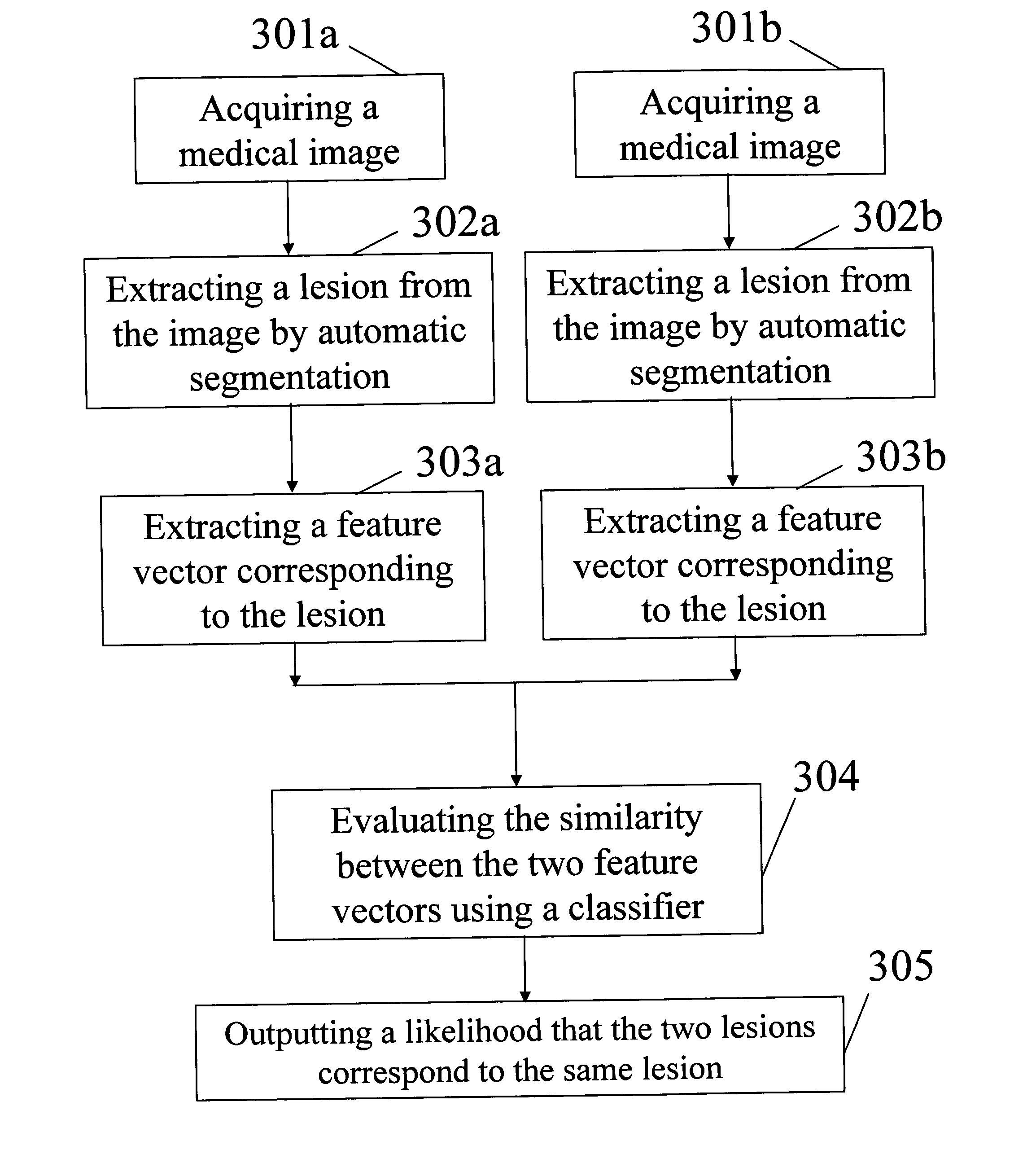
[0065] The aforementioned difficulties are mitigated by using image features associated with the lesions and developing a classification scheme for establishing lesion correspondence. That is, given two breast images, lesions are first automatically segmented from the surrounding breast tissues and a set of features (feature vector) is automatically extracted from each identified lesion. Subsequently, for every two indicated lesions that are not in the same image, a classifier will examine their features and yield the likelihood that the two lesions correspond to the same physical lesion.

[0066] Image features produced by automatic segmentation that have been successful in developing computer-aided diagnosis (CAD) of breast cancers are also useful for the task of lesion matching. An appropriate subset of features useful for discriminating corresponding and non-corresponding lesion pairs will need to be determined. The features will then be used to develop discrimination methods for ...


PUM


Abstract

A method, system, and computer software product for correlating medical images, comprising: obtaining first image data representative of a first medical image including a first abnormality; obtaining second image data representative of a second medical image including a second abnormality; determining at least one feature value for each of the first and second abnormalities using the first and second image data; calculating, based on the determined feature values, a likelihood value indicative of a likelihood that the first and second abnormalities are a same abnormality; and outputting the determined likelihood value.

Description

CROSS-REFERENCE TO RELATED APPLICATIONS [0001] This application claims the benefit under 35 U.S.C. § 119(e) of the filing date of Provisional Application Ser. No. 60 / 544,237, filed Feb. 13, 2004, the contents of which are incorporated herein by reference.BACKGROUND OF THE INVENTION [0002] Field of the Invention [0003] The present invention is directed to feature-based correlation of lesions from multiple images. More precisely, the present invention relates to the correlation of lesions observed on breast images acquired from different modalities, which may include, e.g., mammography, breast sonography, and magnetic resonance imaging (MRI). The present invention also relates to the correlation of lesions observed on breast images acquired from different views, times, or protocols, of a single modality for a given patient. [0004] The present invention also generally relates to computerized techniques for automated analysis of digital images, for example, as disclosed in one or more o...

Claims


Application Information

Patent Timeline
IPC IPC(8): A61B5/05G06T7/00
CPCG06T7/0012G06T7/0022G06T7/0028Y10S128/922G06T2207/10116G06T2207/10132G06T2207/30068G06T7/0034G06T7/97G06T7/33G06T7/35
Inventor GIGER, MARYELLEN L.WEN, HUIHUALAN, LI
Owner UNIVERSITY OF CHICAGO
Learn More