Check patentability & draft patents in minutes with Patsnap Eureka AI!

X-ray computed tomography apparatus

Active Publication Date: 2006-01-26
TOSHIBA MEDICAL SYST CORP
View PDF4 Cites 10 Cited by
  • Summary
  • Abstract
  • Description
  • Claims
  • Application Information

AI Technical Summary

Benefits of technology

[0011] The present invention has been made in consideration of the above situation, and has as its object to provide an X-ray computed tomography apparatus which can shorten the imaging time and reduce the operation load on a person who performs imaging while reducing the exposure to X-rays of a subject to be examined, by allowing to set an image SD for each imaging region in an imaging sequence.

Problems solved by technology

In the conventional system, however, an image SD cannot be set for each imaging region in a sequence.
This increases the time and labor required for imaging.

Method used


Image

  • X-ray computed tomography apparatus
  • X-ray computed tomography apparatus
  • X-ray computed tomography apparatus

Examples


Embodiment Construction

[0021] An embodiment of the present invention will be described with reference to the views of the accompanying drawing. In the following description, the same reference numerals denote constituent elements having almost the same functions and arrangements, and a repetitive description thereof will be made only when required.

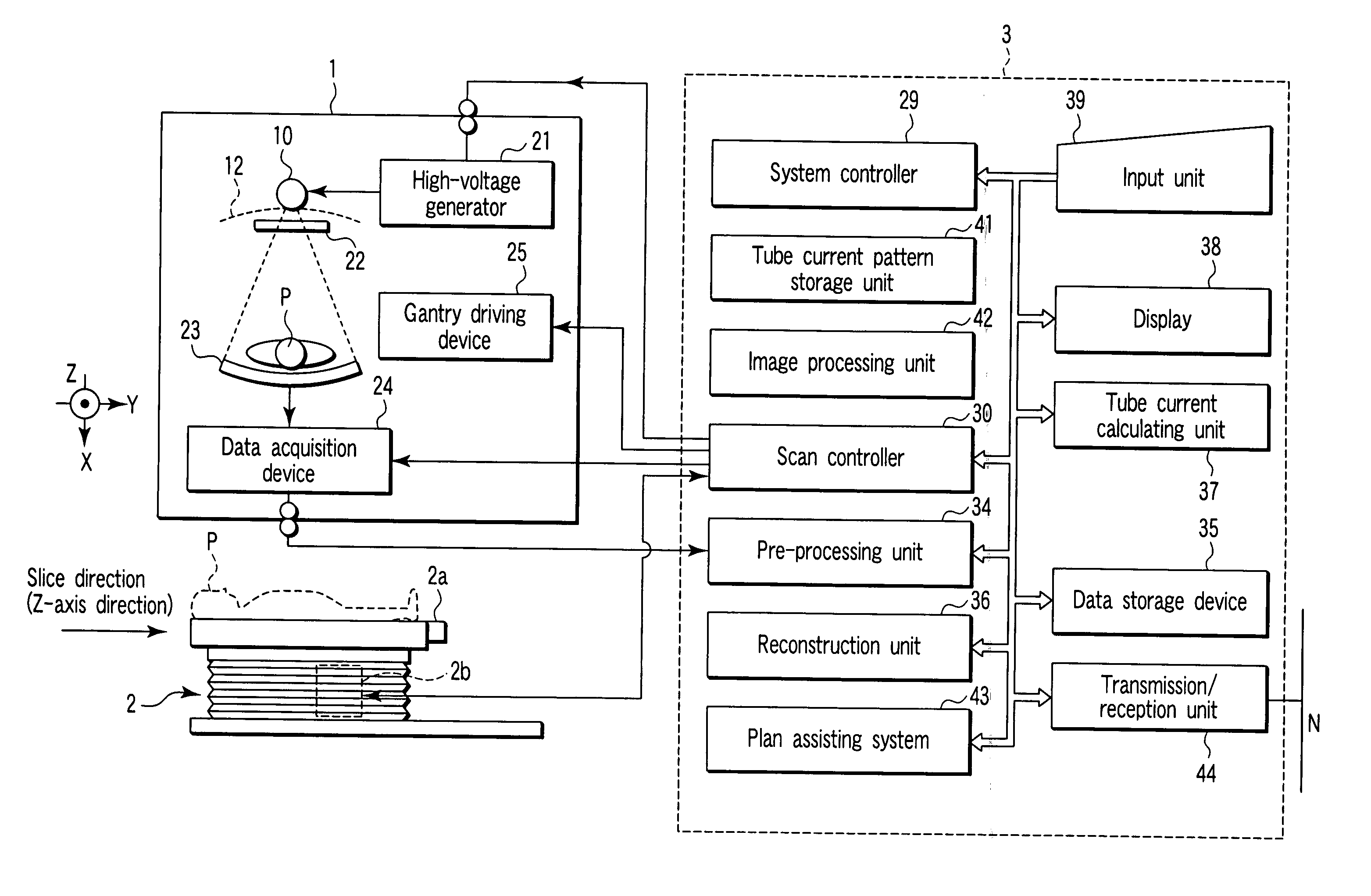
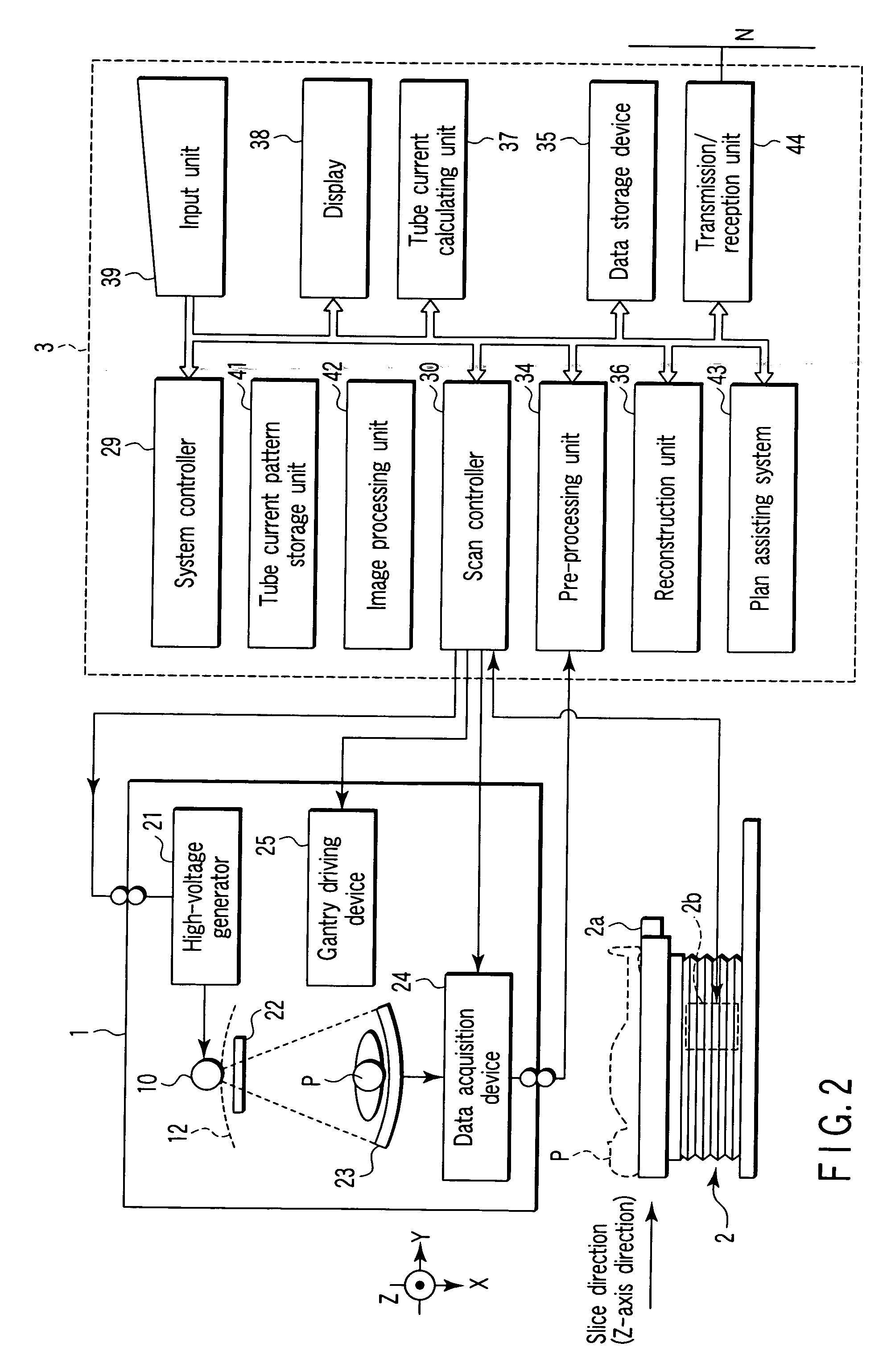
[0022] An embodiment of an X-ray computed tomography apparatus according to the present invention will be described below with reference to the views of the accompanying drawing. Note that X-ray computed tomography apparatuses include various types of apparatuses, e.g., a rotate / rotate-type apparatus in which an X-ray tube and X-ray detector rotate together around a subject to be examined, and a stationary / rotate-type apparatus in which many detection elements arrayed in the form of a ring, and only an X-ray tube rotates around a subject to be examined. The present invention can be applied to either type. In this case, the rotate / rotate type, which is currently...


PUM


Abstract

A plurality of region-specific areas are set on a scanogram, and an image SD value is set for each region-specific area. A tube current calculating unit calculates a tube current value at each position on the basis of a tube current pattern in a tube current pattern storage unit, an image SD value for each region-specific area, and a CT value at each position on the scanogram in each region-specific area. A scan controller controls X-ray emission in accordance with a calculated tube current value at each position.

Description

CROSS-REFERENCE TO RELATED APPLICATIONS [0001] This application is based upon and claims the benefit of priority from prior Japanese Patent Application No. 2004-216308, filed Jul. 23, 2004, the entire contents of which are incorporated herein by reference. BACKGROUND OF THE INVENTION [0002] 1. Field of the Invention [0003] The present invention relates to an X-ray computed tomography apparatus which can set appropriate tube currents (mA) for different regions upon one helical scan by designating an image SD (Standard Deviation) for each region. [0004] 2. Description of the Related Art [0005] As is known, an X-ray computed tomography apparatus is designed to obtain an image (tomographic image) by calculating (reconstructing) the X-ray absorption coefficient of tissue such as an organ on the basis of the amount of X-rays absorbed in a subject to be examined as an index called a CT value with reference to the X-ray absorption coefficient of water. [0006] A reconstructed image inevitabl...

Claims


Application Information

Patent Timeline
IPC IPC(8): H05G1/60A61B6/00G01N23/00G21K1/12
CPCA61B6/032A61B6/027A61B6/542
Inventor NABATAME, TAKEO
Owner TOSHIBA MEDICAL SYST CORP
Features
  • R&D
  • Intellectual Property
  • Life Sciences
  • Materials
  • Tech Scout
  • Unparalleled Data Quality
  • Higher Quality Content
  • 60% Fewer Hallucinations
Learn More