Method and appartus for controlling ultrasound system display

Active Publication Date: 2006-01-26
GENERAL ELECTRIC CO
View PDF29 Cites 102 Cited by
  • Summary
  • Abstract
  • Description
  • Claims
  • Application Information

AI Technical Summary

Benefits of technology

[0004] In one embodiment, a method for operating a medical imaging system is provided. The method includes receiving an image data set of a region of interest in a first dimensional representation, reducing the dimensionality

Problems solved by technology

Large data sets can make managing and manipulating the data hardware intensive, such as requiring large amounts of random access memory (RAM), large disk storage, powerful microprocessors, and image handling chipsets.
The large data sets also may make navigating though the data, locating an object of interest, and displaying a desired view of the object of interest cumbersome and difficult.
Because the physical

Method used


Image

  • Method and appartus for controlling ultrasound system display
  • Method and appartus for controlling ultrasound system display
  • Method and appartus for controlling ultrasound system display

Examples


Embodiment Construction

[0011] Exemplary embodiments of ultrasound systems and methods for controlling such systems are described in detail below. A detailed description of exemplary ultrasound systems will first be provided followed by a detailed description of an embodiment that facilitates managing, navigating, and displaying image data in ultrasound systems.

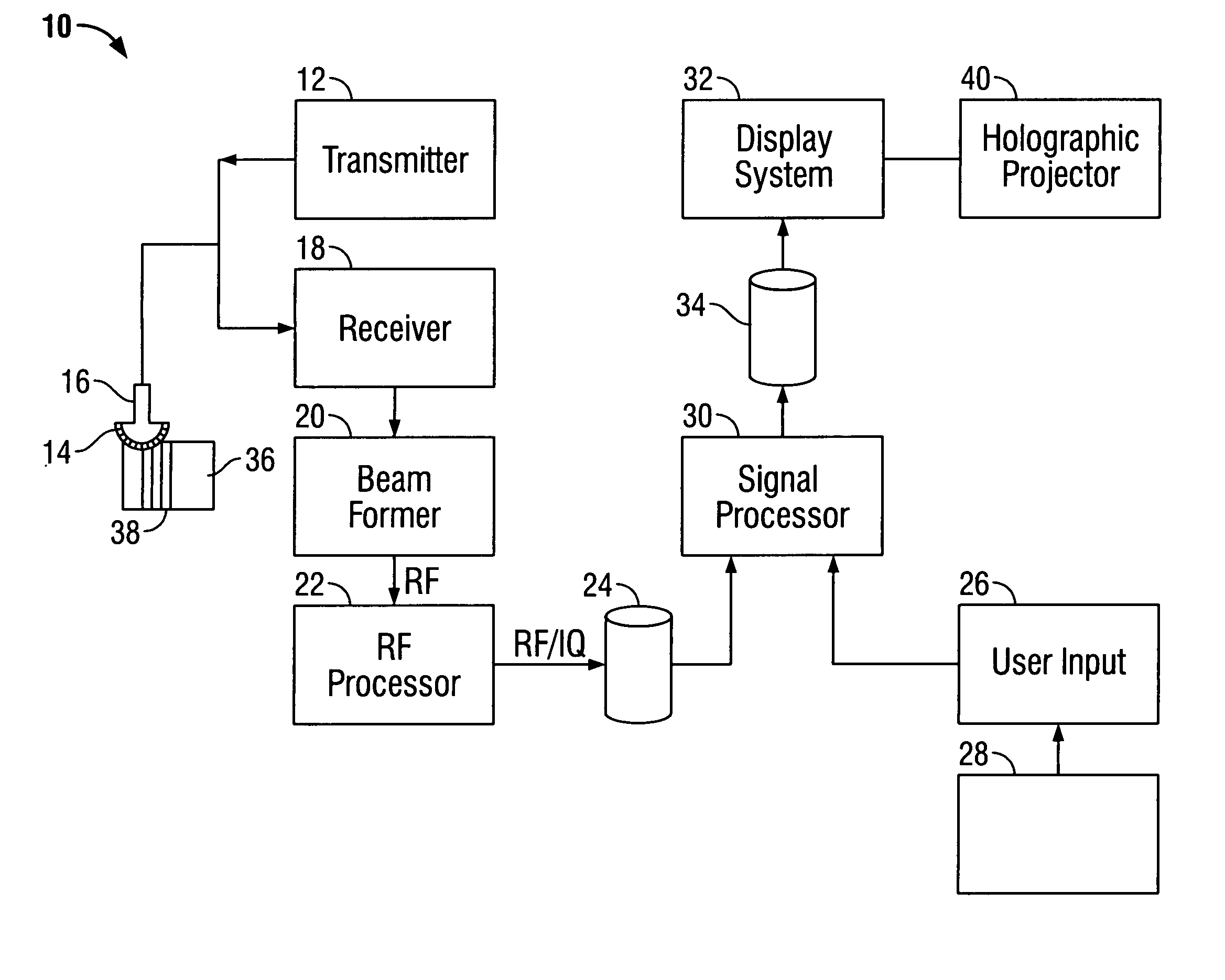
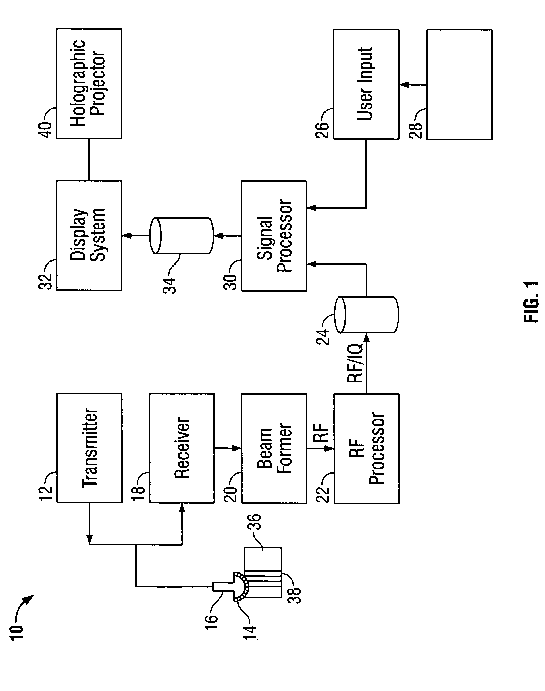
[0012]FIG. 1 is a block diagram of an ultrasound system in accordance with one exemplary embodiment of the present invention. Ultrasound system 10 includes a transmitter 12 that drives transducer elements 14 within a probe 16 to emit pulsed ultrasonic signals into a body. A variety of geometries may be used. The ultrasonic signals are back-scattered from structures in the body, like blood cells or muscular tissue, to produce echoes that return to transducer elements 14. The echoes are received by a receiver 18. The received echoes are provided to a beamformer 20, which performs beamforming and outputs an RF signal. The RF signal is then transmitted...


PUM


Abstract

A method for operating a medical imaging system is provided. The method includes receiving an image data set of a region of interest in a first dimensional representation, reducing the dimensionality of the image data set to a second dimensional representation, selecting a feature of interest in the second dimensional representation, and generating an image of the selected feature in the first dimensional representation.

Description

BACKGROUND OF THE INVENTION [0001] This invention relates generally to ultrasound systems and, more particularly, to methods and devices for controlling imaging in ultrasound systems. [0002] At least some known ultrasound systems, for example, an ultrasound machine or scanner, are capable of performing three dimensional (3D) volume date acquisition and acquisition of the 3D data sets progressing over time (4D). Raw data and processed data of the 3D and 4D data sets may grow exponentially as multiple dimensional data are spanned. Large data sets can make managing and manipulating the data hardware intensive, such as requiring large amounts of random access memory (RAM), large disk storage, powerful microprocessors, and image handling chipsets. The large data sets also may make navigating though the data, locating an object of interest, and displaying a desired view of the object of interest cumbersome and difficult. Because the physical space on the ultrasound machine may be limited,...

Claims


Application Information

Patent Timeline
IPC IPC(8): A61B8/00
CPCA61B8/463A61B8/465A61B8/467G01S15/8993G01S7/52073G01S7/52074G01S7/52084A61B8/483A61B8/466
Inventor MATHEW, PRAKASH PARAYILANGLE, MICHELLE GANIEREMILLER, STEVEN CHARLES
Owner GENERAL ELECTRIC CO
Why Eureka
Try Eureka