System and method for creating magnified images of a microscope slide

a microscope slide and magnifying technology, applied in the field of microscope slide magnifying technology, can solve the problems of insufficient information for the first sequence of images, the inability to capture an entire slide in focus using a fixed vertical, or z-position, and the existing start/stop acquisition system, as described above, to achieve the effect of optimal image quality characteristics

Inactive Publication Date: 2006-03-02
CARL ZEISS MICROSCOPY GMBH
View PDF11 Cites 77 Cited by
  • Summary
  • Abstract
  • Description
  • Claims
  • Application Information

AI Technical Summary

Benefits of technology

[0011] The invention is directed, generally, to a microscopy system for scanning physical sample slides, the physical slides moveable through a microscope optical field of view and focusable through a z position, and relates particularly to a method for creating a digital, virtual slide having optimum image quality characteristics. The method comprises the identification of multiple regions for each physical slide and the definition of at least two focus z positions z1, and z2. Each region of the physical slide is scanned (imaged) at the first z position, so as to produce a first set of digital images of each defined region. Each region of the physical slide is also scanned (imaged) at the second z position, so as to produce a second set of digital images of each defined region. Each image of each set is evaluated against a focus quality metric and, for each region, either the first or second image, corresponding to that region, is selected that exhibits a focus quality metric corresponding to a desired focus quality.

Problems solved by technology

Additionally, the surfaces of many tissue types are uneven and contain local variations that make it difficult to capture an in-focus image of an entire slide using a fixed vertical, or z-position.
In some cases, a first sequence of images does not provide sufficient information to determine a desired focus position.
Existing start / stop acquisition systems, as described above, are relatively slow because the microscope objective lens is often required to perform multiple focus-capture operations for each designated target point on the microscopic slide.
In addition, the field of view of high-power objective lenses is necessarily limited; therefore, the number of points for which desired-focus information is directly obtained may represent a relatively small portion of the entire slide.
First, as described above, the use of a high-power objective lens to obtain desired-focus data for a given target point is relatively slow.
Second, generating a focal map from a limited number of points on the slide can create inaccuracies in the resulting focal map.
Tissue on a slide often does not have a uniform, smooth surface.
Intra-field variations can also cause focus information to be inaccurate.
Even when the focus information is accurate, the mechanical nature of the microscope apparatus can cause a scan to produce an out-of-focus image due to mechanical problems; such as small motions or vibrations of the apparatus, incorrect calibration, etc.

Method used


Image

  • System and method for creating magnified images of a microscope slide
  • System and method for creating magnified images of a microscope slide
  • System and method for creating magnified images of a microscope slide

Examples


Embodiment Construction

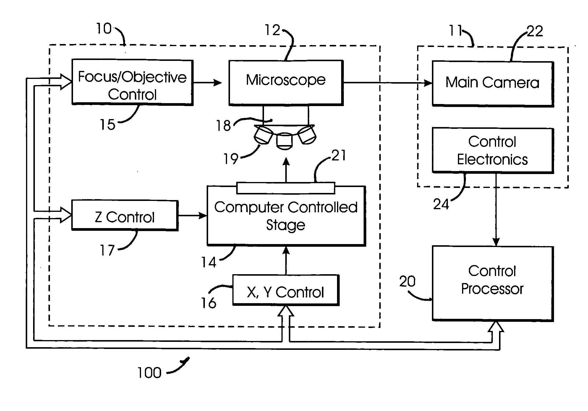
[0030] A virtual microscope slide typically comprises digital data representing a magnified image of all, or a portion of, a microscope slide. Because the virtual slide is in digital form, it can easily be stored on a digital storage medium, such as in the form of an image file in a computer memory, or on a disk drive, a CD ROM, or the like, and can easily be transmitted as a digital file over a communication network, such as the Internet, an intranet, etc., to a suitable viewer at a remote location.

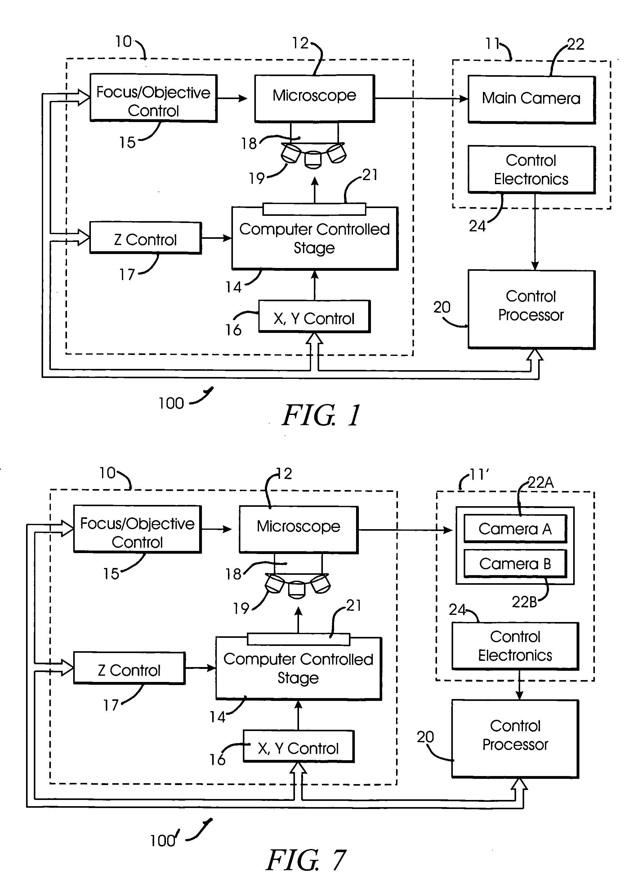
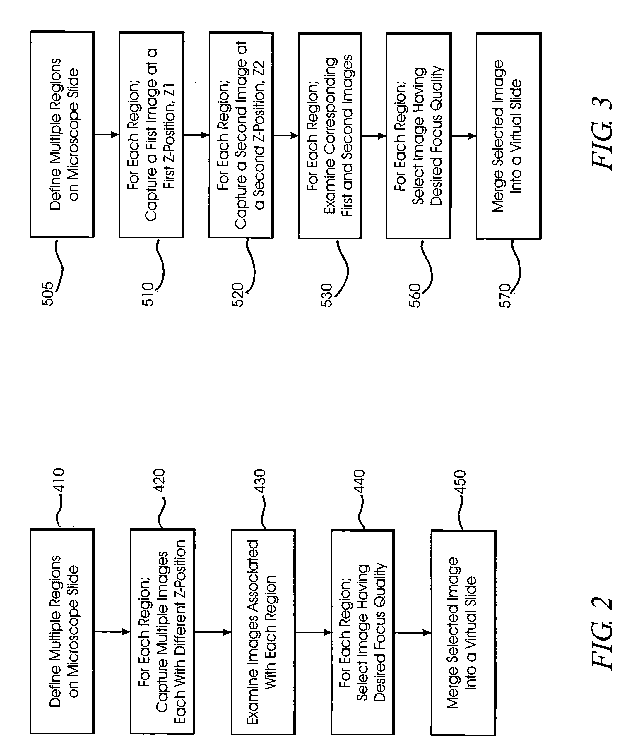
[0031] The various embodiments, described herein, provide improved systems and methods for obtaining in-focus images of desired portions of a microscope slide and further provide improved systems and methods for combining the in-focus images in order to create a complete virtual slide. In one aspect of a an exemplary embodiment of the invention, multiple regions of a microscope slide are defined, multiple images of each region are captured and, for each defined region, an image having a...


PUM


Abstract

A method and system for creating a digital, virtual slide having optimum image quality characteristics. Multiple regions of a physical slide are identified as well as at least two focus z-positions z1, and z2. Each region of the physical slide is scanned (imaged) at the first z position, so as to produce a first set of digital images of each defined region. Each region of the physical slide is also scanned (imaged) at the second z position, so as to produce a second set of digital images of each defined region. Each image of each set is evaluated against a focus quality metric and, for each region, either the first or second image, corresponding to that region, is selected that exhibits a focus quality metric corresponding to a desired focus quality. These images are then merged into a digital virtual slide. Additional focus z-positions may be included, and the multiple z-positions may be scanned seriatim, sequentially, and/or in overlapping fashion.

Description

CROSS REFERENCE TO RELATED APPLICATIONS [0001] The present application is related to and takes priority from U.S. Provisional Applications Ser. Nos. 60 / 605,835, and 60 / 609,961, filed Aug. 31, 2004 and Sep. 15, 2004, respectively, and is further related to co-pending U.S. patent application entitled SYSTEMS AND METHODS FOR STITCHING IMAGE BLOCKS TO CREATE SEAMLESS MAGNIFIED IMAGES OF A MICROSCOPE SLIDE, all commonly owned by the assignee of the present application, the entire contents of which are expressly incorporated herein by reference.FIELD OF THE INVENTION [0002] The invention relates generally to a system and method for obtaining image data for use in creating virtual microscope slides, and more particularly, to a method and system for obtaining magnified images of a microscope slide. BACKGROUND OF THE INVENTION [0003] A virtual microscope slide typically comprises digital data representing a magnified image of a microscope slide. Because the virtual slide is in digital form, ...

Claims


Application Information

Patent Timeline
Patent Type & Authority Applications(United States)
IPC IPC(8): G03B13/32
CPCG02B21/365G02B21/367G06T2207/30004G06T7/0026G06T2200/32G02B27/0075G06T7/32
Inventor ZEINEH, JACK A.DONG, RUI-TAO
Owner CARL ZEISS MICROSCOPY GMBH
Why Eureka
Try Eureka