Nasal oral respiratory interface

a nasal cavity and nasal tube technology, applied in the direction of respirators, respiratory apparatus, catheters, etc., can solve the problems of allowing the tube to move, poor fixation of adhesive tape, and damage to the larynx, so as to prevent movement or kinking of the tracheal tube, and ensure the attachment of the device, not to negatively affect the growth and development of the oral cavity

Inactive Publication Date: 2006-04-20
PENN STATE RES FOUND
View PDF54 Cites 85 Cited by
  • Summary
  • Abstract
  • Description
  • Claims
  • Application Information

AI Technical Summary

Benefits of technology

[0017] One advantage of the present invention is that it provides an interface for the attachment of an assortment of medical devices that improves upon the limitations of current attachment methods. In particular, it avoids the use of adhesive tape applied to the face, while providing a more stable and a secure attachment of these devices.
[0018] Another advantage of the present invention is that it provides a stable tracheal tube holder and tracheal tube stent, thereby maintaining the angle the tube enters the mouth, and preventing movement or kinking of the tracheal tube. In this manner, the tracheal tube will not impinge upon the infants lip or palate, and therefore, will not negatively impact upon oral-facial growth and development.
[0019] Still another advantage of the present invention is that because the endotracheal tube is better stabilized, the risk of accidental extubation is significantly reduced. This increase in stabilization also prevents tube movement during mechanical ventilation, thereby lessening the potential for patient discomfort, desaturation spells and laryngal trauma.
[0020] Yet another advantage of the present invention is that the transverse stent is stronger and more resistant to bending and buckling than known stabilization systems. By providing the ability to easily add additional anchorage, heavier medical devices, such as nasal CPAP cannula, can be fully supported, thereby reducing patient discomfort associated with those heavier devices.

Problems solved by technology

Serious complications can result from accidental extubation (dislodgment) of these tubes, including acute hypoxia, bradycardia, and long term laryngaltracheal damage from reintubation.
However, adhesive tape stabilization methods provide poor fixation, allowing the tube to move when the skin is stretched.
The tape obstructs the face, has the potential to obstruct the nasal openings, and loosens with time.
Tape is difficult to remove and reapply when adjustments to the tubes are required, often causing injuries to the skin by stripping away the epidermis, especially in premature infants.
Tape adhesion methods have a further disadvantage, in that nasal and oral secretions are absorbed by the tape, causing the tape to lose adhesion and loosen, while contaminating the tape with microbial organisms that could colonize the patient and gain direct access to the lung via the endotracheal tube and cause pneumonia
As a result of this pulling force, an improperly stabilized tube may cause pain and discomfort if the tube slides up and down within the trachea, especially if it hits against the bifurcation of the trachea.
Moreover if the tube protruding outside the patient kinks, gas flow will be obstructed.
Such movement of the endotracheal tube in the small premature infant is a major clinical problem.
With the exception of the bar, these known stabilization attempts have covered the patient's mouth and portions of the tube, making oral hygiene and tube visualization difficult.
These devices further require the endotracheal tube be placed within the device prior to intubation, thereby blocking the clinician's visualization of the trachea and increasing the difficulty of successfully placing the tube in its desired location, making the device potentially dangerous.
Additionally, the tube can rotate around the bar, allowing it to kink in the patient's larynx, restricting air flow, or placing extended tube pressure on the palate, causing interference with normal palatal development.
However, these plates interfere with oral hygiene and are difficult and clumsy to place properly.

Method used


Image

  • Nasal oral respiratory interface
  • Nasal oral respiratory interface
  • Nasal oral respiratory interface

Examples


Embodiment Construction

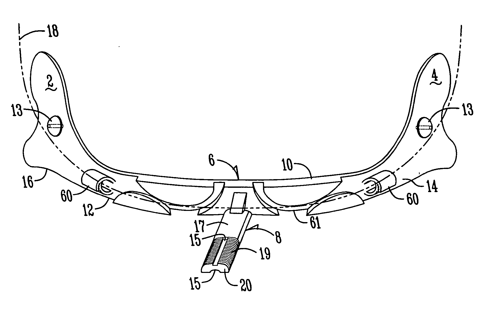
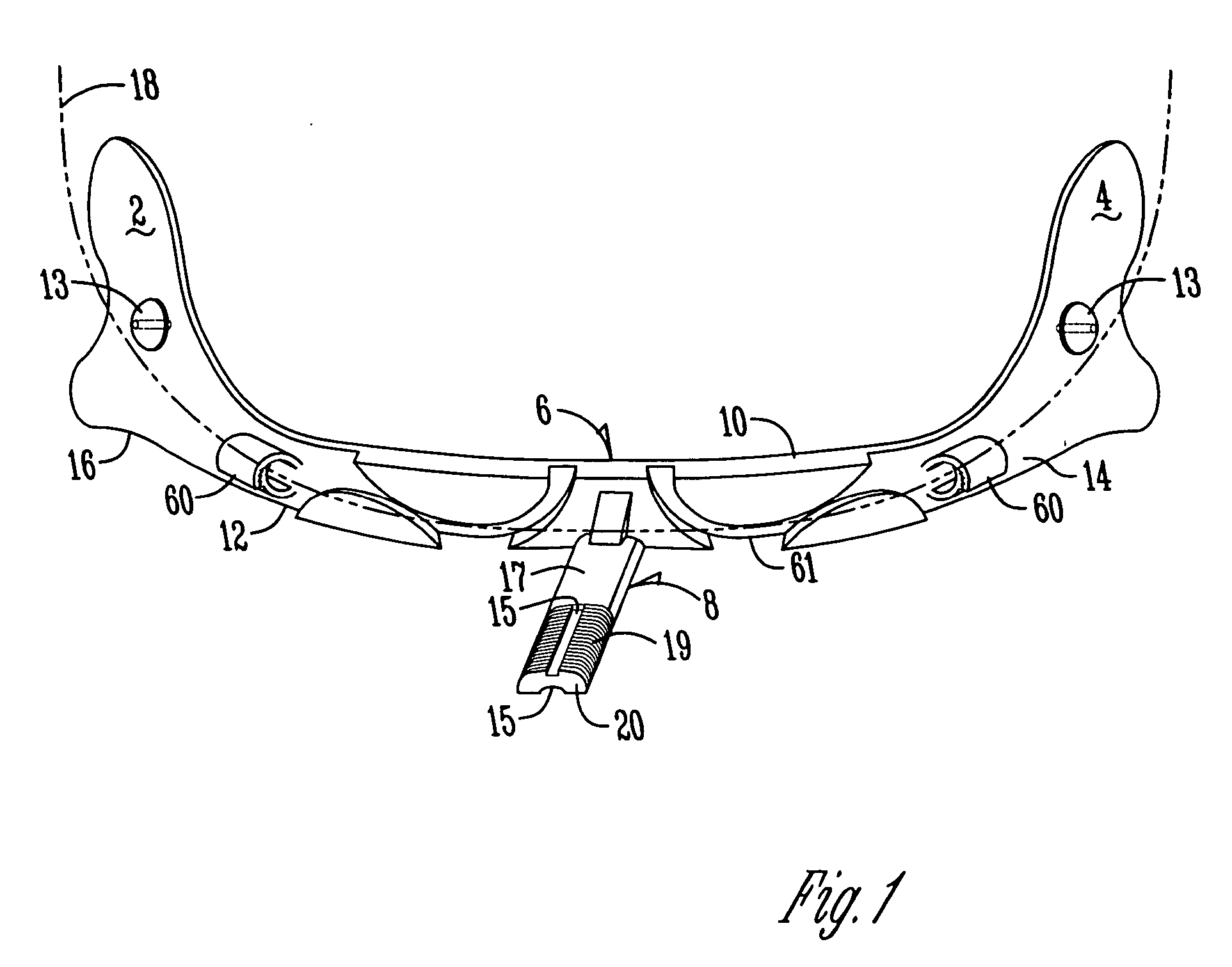
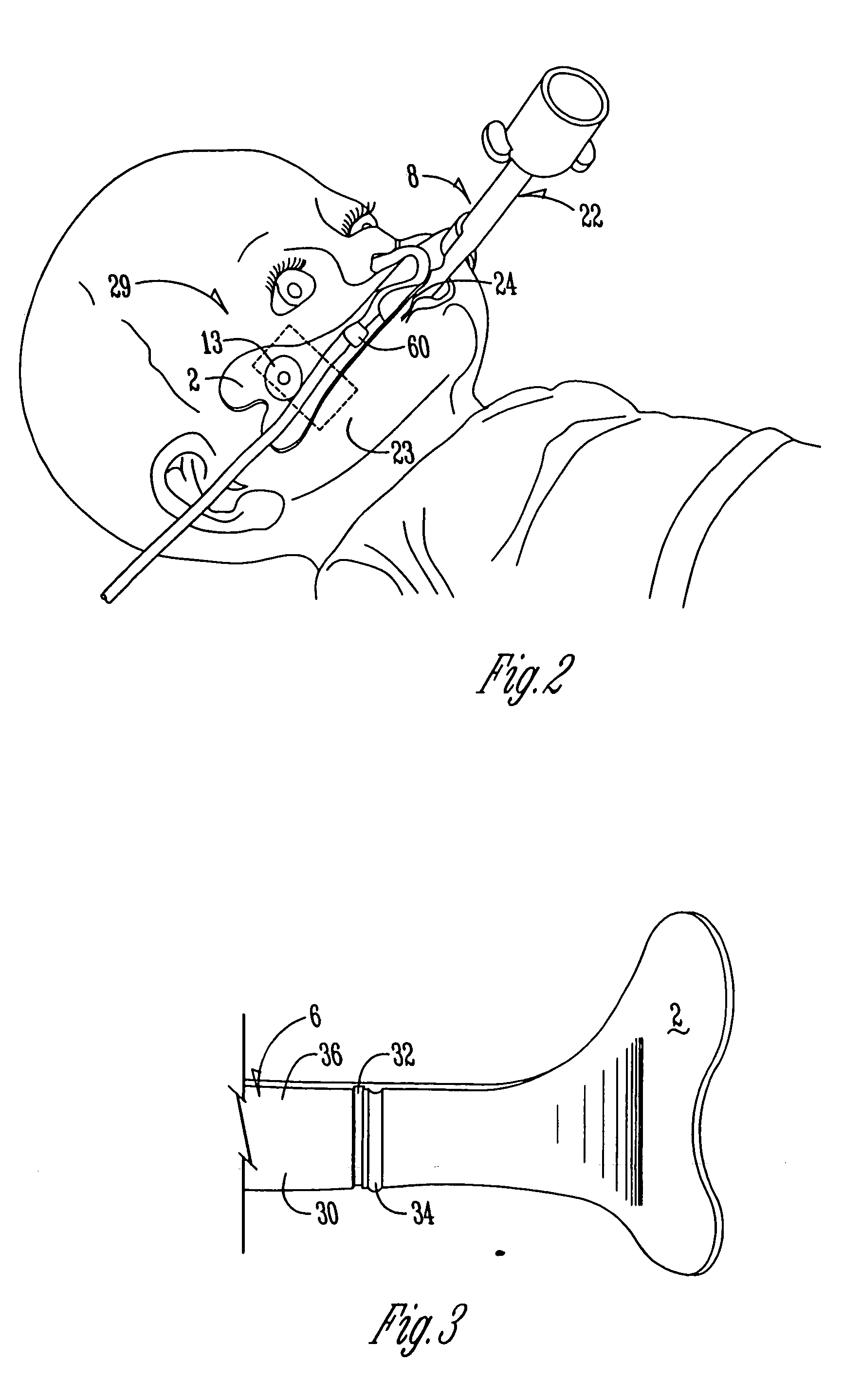
[0031] The present invention sets forth a method and an apparatus for stabilizing at least one medical device entering at least one facial cavity. Such medical devices may include, for example, endotracheal tubes, nasotracheal tubes, nasalgastric tubes, naso or orojejunal tubes, nasal thermistors, nasal pneumotachometers, nasal capnographs, nasal masks, oxygen delivery tubing and nasal CPAP delivery systems, although the present invention will stabilize any device entering or covering the mouth or nose. For the purposes of this disclosure, the term “tube” is meant to include all such medical devices entering the mouth or nose, and that the present invention is not limited to stabilizing medical devices tubular in shape. Likewise, it should be understood that oral insertion, nasal insertion or both simultaneously will be stabilized equally well.

[0032] As shown in FIG. 1, the present invention comprises a first 2 and second 4 facial interface positioned on opposite terminal ends of a...


PUM


Abstract

The present invention provides an apparatus and a method for stabilizing at least one medical device such as a tracheal or gastric tube entering or covering at least one facial cavity such as the mouth and / or nose, taking advantage of a stabilization stent to secure to a patient's face the tracheal or gastric tube without the need to apply tape to the face, while maintaining visibility of tube markings, preventing kinking of the tube, and keeping the infant's face visible to parents and caregivers. In one form, the invention comprises a transverse stent comprising a superior border opposed to an inferior border, an inner surface and an outer surface. A first facial interface is attached to a first terminal end of the transverse stent, and positioned to adhesively adhere and mechanically clasp to a first side of a patient's face. A second facial interface is attached to a second terminal end of the transverse stent and positioned to adhesively adhere and mechanically clasp to a second side of a patient's face. A docking platform attached to the outer surface of the transverse stent supports the medical device. In a different form, the invention also includes gutters positioned on the inner surface of the respective first and second terminal end approximate to the respective first and second facial interface to channel oral fluids away from the respective first and second facial interface. The outer surface contains channels for containing and directing tubing, and fixaton points for attaching anchoring measures such as chin or head straps. The core of the transverse stent is hollowed to permit gas flow.

Description

CROSS REFERENCE TO RELATED APPLICATIONS [0001] This application claims benefit of U.S. Provisional Application Nos. 60 / 186,859 filed Mar. 3, 2000 and 60 / 201,935 filed May 5, 2000.FIELD OF THE INVENTION [0002] This invention relates generally to stabilization of medical devices, and more particularly to stabilization of devices that enter or cover the nose and / or mouth. Although most useful in a neonatal intensive care unit (NICU) setting, the advantages of the present invention are applicable to any patient age group or setting requiring such stabilization. BACKGROUND OF THE INVENTION [0003] Infants requiring mechanical ventilation (assisted breathing) are commonly treated by endotracheal intubation, wherein a flexible tube is inserted into the mouth, down the throat and through the lumen of the trachea a predetermined distance, to deliver oxygen to the lungs. In addition, flexible feeding tubes may also be inserted through the nose or mouth, passing through the esophagus, and termi...

Claims


Application Information

Patent Timeline
Patent Type & Authority Applications(United States)
IPC IPC(8): A62B9/06A61M16/00A61M16/04A61M16/06
CPCA61M16/0488A61M16/0497A61M16/0666A61M16/0683A61M2025/022A61M2025/0226A61M16/0672A61M16/0688A61M16/0694
Inventor PALMER, CHARLES
Owner PENN STATE RES FOUND
Why Eureka
Try Eureka