Method for genome-wide analysis of palindrome formation and uses thereof

a genome-wide analysis and palindrome technology, applied in the field of genome-wide analysis of palindrome formation, can solve the problems of limited examples, dna preparation, and inability to detect dsb repair,

Inactive Publication Date: 2006-04-27
FRED HUTCHINSON CANCER RES CENT
View PDF6 Cites 5 Cited by
  • Summary
  • Abstract
  • Description
  • Claims
  • Application Information

AI Technical Summary

Benefits of technology

[0008] A genome-wide method for identifying a region of genomic DNA comprising a DNA palindrome is disclosed. The method generally comprises incubating isolated fragmented total genomic DNA under conditions conducive to snap back DNA formation and not inter-molecular hybridization, the snap back DNA containing the DNA palindrome; isolating the snap back DNA; and identifying the regions of the genomic DNA comprising the snap back DNA to identify those regions of the genomic DNA c...

Problems solved by technology

This indicates that improper DSB repair also could trigger the BFB cycle for further chromosome aberrations.
However, our molecular analysis of the structure of amplified loci in cancer cells has been ...

Method used


Image

  • Method for genome-wide analysis of palindrome formation and uses thereof
  • Method for genome-wide analysis of palindrome formation and uses thereof
  • Method for genome-wide analysis of palindrome formation and uses thereof

Examples


example 1

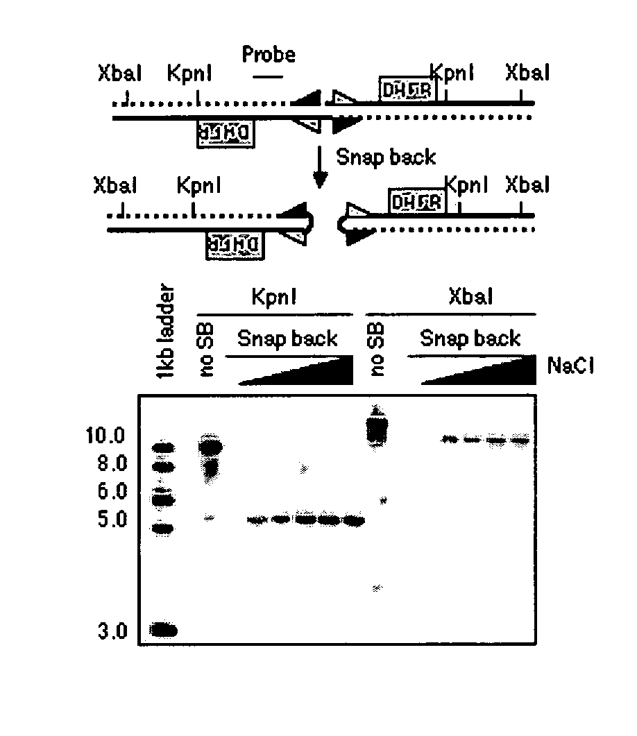
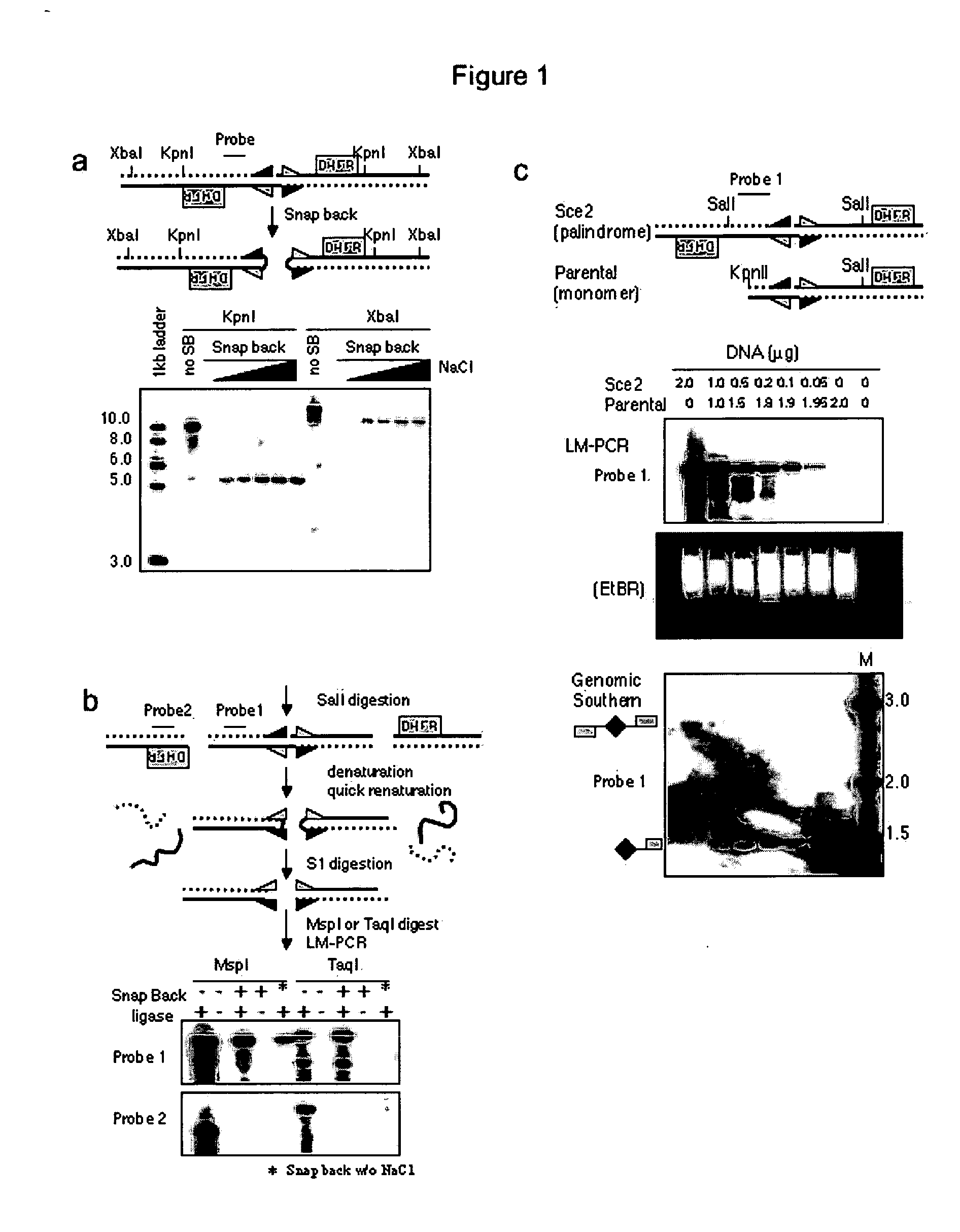
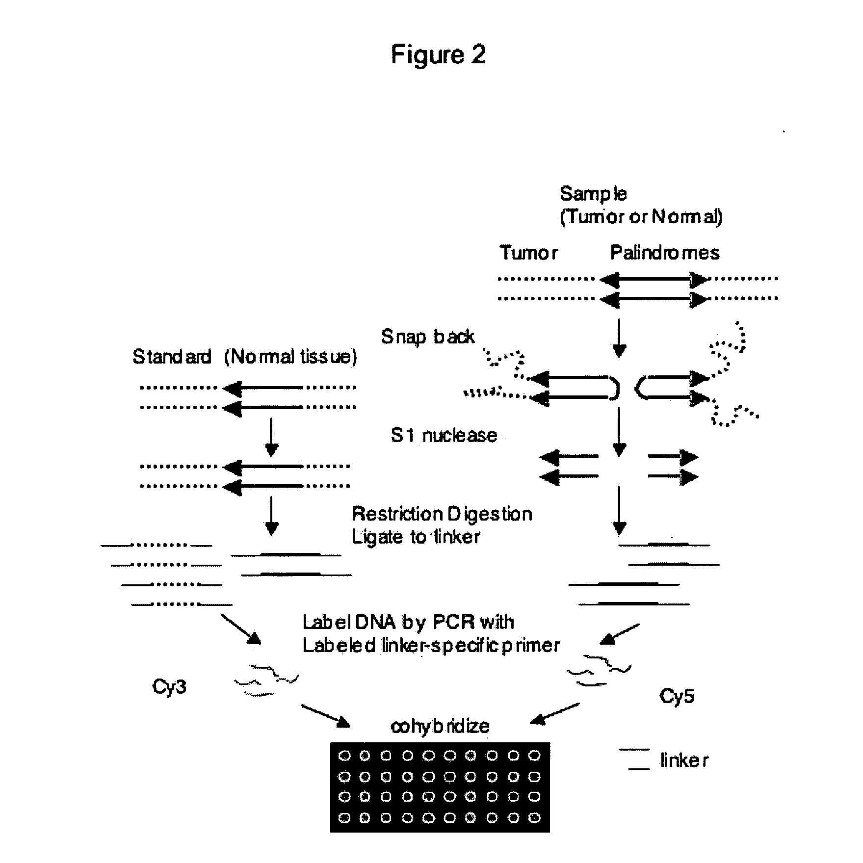
[0026] The following example describes the process for genome-wide assessment of palindrome formation.

Methods

Cell Lines and Cancer Tissues

[0027] D79IR-8 and D79IR-8-Sce 2 cells were previously described (Tanaka et al., Proc. Natl. Acad. Sci. USA 99:8772-8777 (2002)). Colo320DM and RD were obtained from American Type Culture Collection. MCF7 and AG1113215 were from the University of Washington. Skin biopsy derived fibroblasts HDF1 and HDF3 were obtained from the University of Washington and human foreskin fibroblasts HFF2 from the Fred Hutchinson Cancer Research Center (FHCRC) as anonymous cell lines. DNA samples stripped of identifying information from five primary medulloblastomas were provided by the FHCRC. All samples were obtained after FHCRC Institutional Review Board review and approval for use of anonymous human DNA samples and human cell lines.

Linkers and Oligos

[0028] Oligonucleotides were synthesized by QIAGEN Genomics. For ligation mediated PCR, two oligonucleotide...

example 2

[0049] The following example demonstrates the use of ligation-mediated PCR to isolated a DNA fragment enriched in unmethylated CpG islands in a mammalian cell. A schematic of the process is provided as FIG. 8A. The methods for

[0050] Briefly, mouse genomic DNA was digested with a methylation sensitive restriction enzyme (for example, HpaII). The MspI linkers used above in Example 1 were used to ligate the HpaII fragments. The ligated DNA was amplified by PCR using the MspI primer from Example 1 (SEQ ID NO: 6). The method resulted in the specific amplification of HpaII digested genomic DNA of less than 500 base pairs. (FIG. 8B). Random cloning and sequencing of the PCR products revealed that more than 50% of clones were at the CpG islands as defined using stringent criteria. (Takai and Jones, Proc. Natl. Acad. Sci USA 99:3740-3745 (2002); incorporated herein by reference). In contrast, amplification of DNA digested with methylation-resistant isoschizomer MspI gave no clones near CpG ...


PUM

PropertyMeasurementUnit
Fractionaaaaaaaaaa
Fractionaaaaaaaaaa
Fractionaaaaaaaaaa

Abstract

The present invention provides a method for rapidly detecting the genome-wide presence of palindrome formation. The method has demonstrated that somatic palindromes occur frequently and are widespread in human cancers. Individual tumor types have a characteristic non-random distribution of palindromes in their genome and a small subset of the palindromic loci are associate with gene amplification. The disclosed method can be used to define the plurality of genomic DNA palindromes associated with various tumor types and can provide methods for the classification of tumors, and the diagnosis, early detection of cancer as well as the monitoring of disease recurrence and assessment of residual disease.

Description

CROSS-REFERENCES TO RELATED APPLICATIONS [0001] The present application claims priority to U.S. Provisional Patent Application No. 60 / 575,331, filed May 28, 2004, the entire disclosure of which is incorporated by reference herein.STATEMENT AS TO RIGHTS TO INVENTIONS MADE UNDER FEDERALLY SPONSORED RESEARCH AND DEVELOPMENT [0002] Aspects of the present invention were conducted with funding provided by the National Institutes of Health under Grant Nos. R01AR 045113 and R01GM 26210. The Government may have certain to rights in the claimed invention.BACKGROUND OF THE INVENTION [0003] Cancer is a disease of impaired genetic integrity. In most cases disturbed genetic integrity is observed at the chromosome level and include a configuration called anaphase bridges, which are most likely derived from dicentric or ring chromosomes segregating into two different daughter cells in the process of the breakage-fusion-bridge (BFB) cycle. The BFB cycles have been shown to generate large DNA palindr...

Claims


Application Information

Patent Timeline
IPC IPC(8): C12Q1/68
CPCC12N15/1093
Inventor TAPSCOTT, STEPHEN J.TANAKA, HISASHIYAO, MENG-CHAO
Owner FRED HUTCHINSON CANCER RES CENT
Why Eureka
Try Eureka