System, imaging suite, and method for using an electro-pneumatic insufflator for magnetic resonance imaging

a magnetic resonance imaging and insufflator technology, applied in medical insufflators, diagnostics, medical science, etc., can solve the problems of early detection, invisible blood to the naked eye, and only detectable microscopically

Inactive Publication Date: 2006-05-11
BRACCO DIAGNOSTICS
View PDF76 Cites 26 Cited by
  • Summary
  • Abstract
  • Description
  • Claims
  • Application Information

AI Technical Summary

Benefits of technology

[0015] According to yet another alternative embodiment of the present invention, a method is provided for using an electro-pneumatic insufflator for magnetic resonance imaging based virtual colonoscopy. Embodiments of the present invention may help provide an improved system and method for using an electro-pneumatic insufflator for magnetic resonance imaging based virtual colonoscopy. As such, the system and method of embodiments of the present invention may solve many of the problems identified by prior techniques and provide additional advantages.

Problems solved by technology

Polyps sometimes bleed, and there may be some noticeable rectal bleeding that leads to early detection of precancerous growths.
However, most of the time, this blood is invisible to the naked eye and is only detectable microscopically.
Currently, the practice of using an electromechanical insufflator to comfortably control distension of the colon with carbon dioxide for radiographic imaging of the colon, typically referred to as virtual colonoscopy, is limited to computed tomography (CT).
Because of the power of these magnets, a room containing an MRI scanner can be very dangerous if precautions are not taken.
Because of the power of MRI magnets, severe safety and functional implications would result if a clinician used an insufflator in an MRI imaging suite.
Introducing these components in direct proximity to the strong magnetic field of the magnetic resonance (MR) scanner could result in a catastrophic interaction.
While it may be theoretically possible to substitute non-ferromagnetic materials for a number of the ferromagnetic materials of the current system, it would require a complete redesign of the pneumatic technology, which would be expensive and time-consuming.
Even if it were possible to replace all ferromagnetic materials in the electromagnetic valves, the introduction of any transient electrical signal in a shielded MR scanner room to control the resultant pneumatic system could potentially interfere with the RF signals generated within the MR scanner.

Method used


Image

  • System, imaging suite, and method for using an electro-pneumatic insufflator for magnetic resonance imaging
  • System, imaging suite, and method for using an electro-pneumatic insufflator for magnetic resonance imaging
  • System, imaging suite, and method for using an electro-pneumatic insufflator for magnetic resonance imaging

Examples


examples

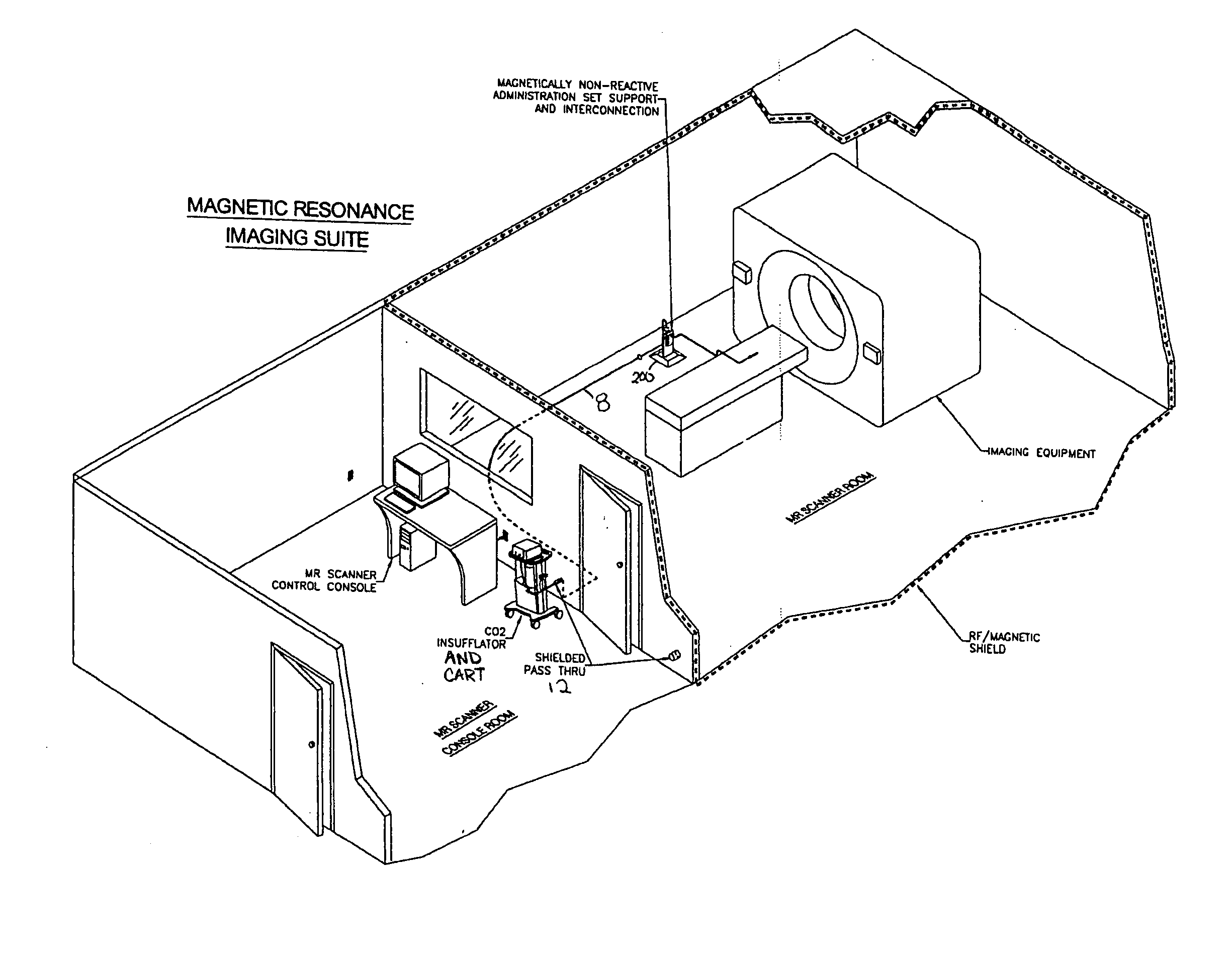
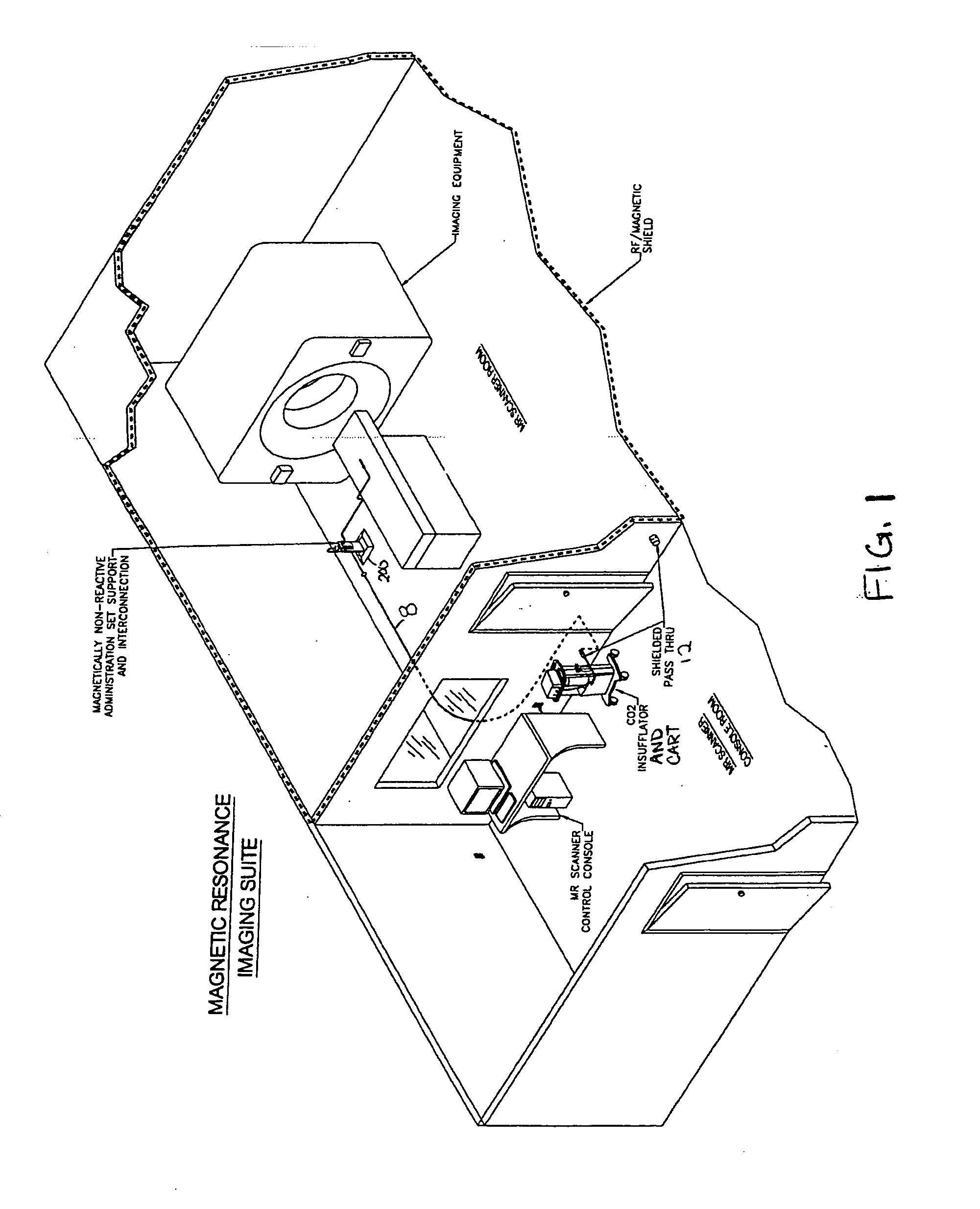
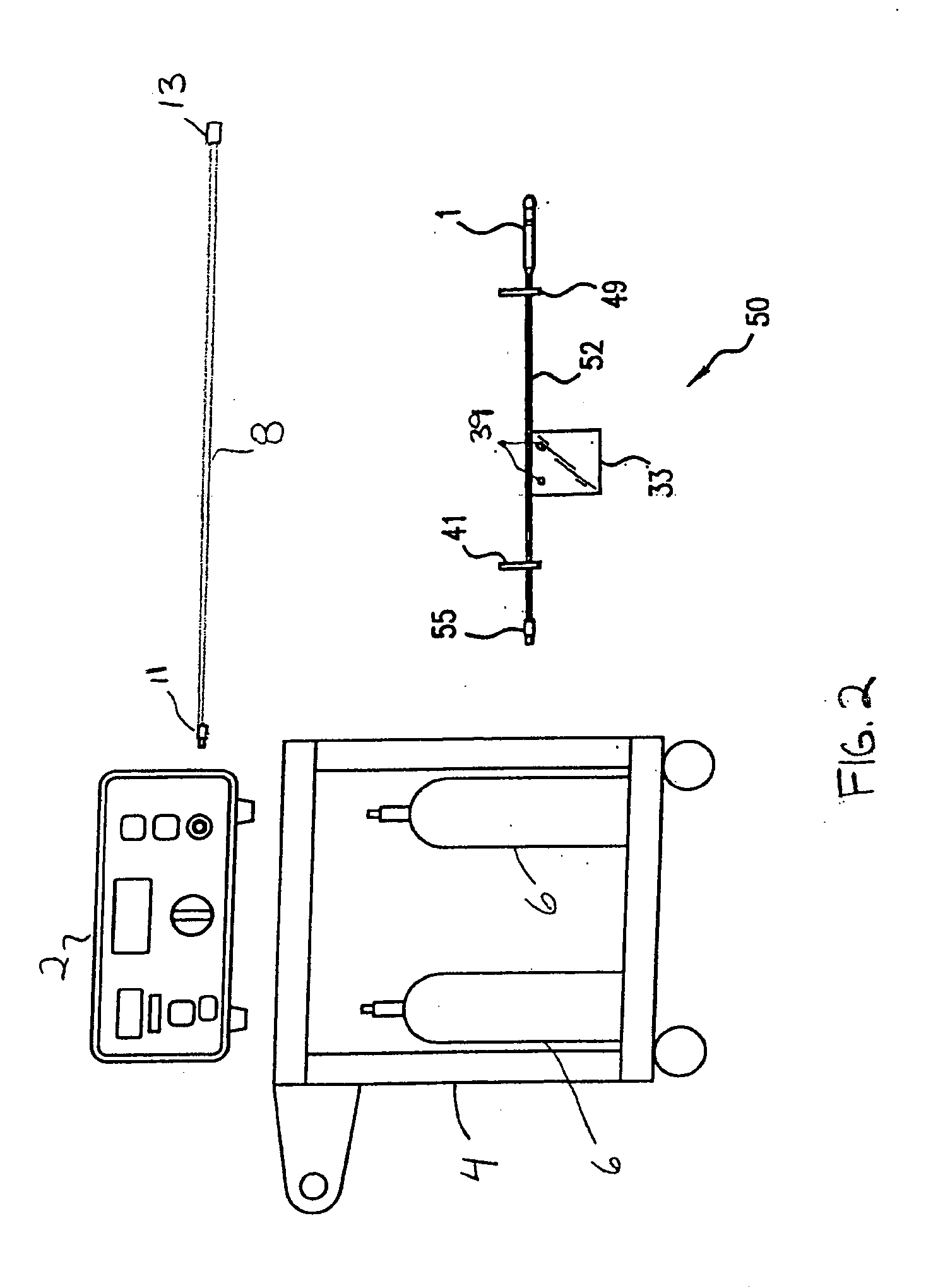
[0057] The following example details a feasibility assessment that was performed pertaining to the potential use of a PROTOCO2L™ Colon Insufflator System in an MRI environment for an MR colonography study. The assessment was limited to comparative measurements of the flow and pressure performance of the PROTOCO2L™ Insufflator and associated devices under experimental conditions first representing CT colonography and secondly representing MR colonography.

[0058] The first experimental set-up representing CT colonography included a measured in-vitro fill into a 3 Liter collapsible container through the standard administration set connected to the PROTOCO2L™ Insufflator. The second experimental set-up representing MR Colonography included a measured in-vitro fill into the same 3 Liter collapsible container through PROTOCO2L™ Administration Set which in turn was connected to a 15.2 meter (50 ft.) electromagnetically inactive connection tube of like diameter to that of the PROTOCO2L™ Adm...


PUM


Abstract

Embodiments of the invention provide a system and method for using an electro-pneumatic insufflator for magnetic resonance imaging, such as for virtual colonoscopy. Embodiments of the invention further provide a MRI imaging suite and a method for distension of a body part to be imaged. As an alternative to the complete redesign of an electromagnetic insufflator, embodiments of the current invention utilize a standard electro-pneumatic insufflator that operates in a location impervious to electromagnetic radiation using an electromagnetically inactive connection tube.

Description

CROSS REFERENCE TO RELATED APPLICATIONS [0001] This application claims priority from U.S. Provisional Application No. 60 / 638,643, filed Dec. 22, 2004 and U.S. Utility application Ser. No. 10 / 497,625; filed Jun. 3, 2004, both of which are incorporated herein by reference in their entirety.FIELD OF THE INVENTION [0002] The invention relates to a suite, system, kit and methodology associated with using an electro-pneumatic insufflator with Magnetic Resonance Imaging (MRI). BACKGROUND OF THE INVENTION [0003] Colorectal cancer, cancer of the large intestine and rectum, is second only to lung cancer in the amount of cancer deaths caused each year. Approximately 5% of all people will develop colorectal cancer within their lifetime. As is true with many other cancers, early detection of colon cancer or its precursors greatly increases chances of survival. [0004] Precancerous polyps begin to form in the colon when cells in the lining of the intestine mutate and begin dividing rapidly. If lef...

Claims


Application Information

Patent Timeline
Patent Type & Authority Applications(United States)
IPC IPC(8): A61B5/05
CPCA61B5/055A61B5/411A61M13/003A61M2202/0225G01R33/28A61B5/004A61B5/0046A61B5/0555
Inventor WILLIAMS, ROBERT C. JR.
Owner BRACCO DIAGNOSTICS
Why Eureka
Try Eureka