Method and system for computer aided detection (cad) cued reading of medical images

a technology medical images, applied in the field of computer aided detection (cad) of abnormalities in xray films, can solve the problems of human and cad results disagreeing, unsettling for physicians, and inability to mark a significant percentage of regions by computer, so as to improve visual diagnosis and reduce false positives

Inactive Publication Date: 2006-06-08
CARESTREAM HEALTH INC
View PDF6 Cites 41 Cited by
  • Summary
  • Abstract
  • Description
  • Claims
  • Application Information

AI Technical Summary

Benefits of technology

[0009] In one aspect of the present invention there is provided a method for displaying results of a computer aided detection (CAD) analysis of a digital or digitized image of a tissue for increasing the accuracy of diagnosis by a user / physician. The method comprises analyzing a digital image of an X-ray film using CAD analysis to produce CAD results, the CAD analysis being capable of detecting regions of the digital image having a greater than zero probability for the presence of one or more abnormalities, and generating one or more coded descriptors of the CAD results, the coded descriptors being associated with the regions detected by CAD analysis and providing information on one or more criteria and / or features used in the CAD analysis. In this manner, when the CAD results are displayed with the associated coded descriptors, the user may evaluate the criteria that were used by the CAD algorithm to arrive at an identification of an abnormal region to enhance the accuracy of his / her diagnosis.
[0012] The use of the coded descriptors advantageously improves visual diagnosis by helping in validating the final result of the analysis, that is, the determination of whether a suspicious region is indeed abnormal (cancerous) and by providing the basis for the CAD analysis that can be compared to the basis of the diagnosis established by the practitioner. The method of the present invention thereby contributes to lower the number of false positives or false negatives associated with X-ray analysis.

Problems solved by technology

It can be very unsettling for the physician if the reason for the discrepancy cannot be easily determined.
The trade-off is that this results in the computer not marking a significant percentage of regions that are potentially cancerous (false-negatives).
A major downside to this approach is that it requires the physician to read each image twice: once with the CAD marks and once without.
A second downside is that once the physician views the CAD results, the human and CAD results may disagree.
Even though some systems may display the CAD results with a probability or likelihood that a suspicious area is cancerous (see for example Zheng et al.
2001, Radiology, 221(3):633-640 and U.S. Patent Applications No. 20020097902, and 20020076091), the CAD results displayed by the computer remain of a “pass / fail” nature and the physician may often struggle in determining the underlying reason for the discrepancy.

Method used


Image

  • Method and system for computer aided detection (cad) cued reading of medical images
  • Method and system for computer aided detection (cad) cued reading of medical images
  • Method and system for computer aided detection (cad) cued reading of medical images

Examples


Embodiment Construction

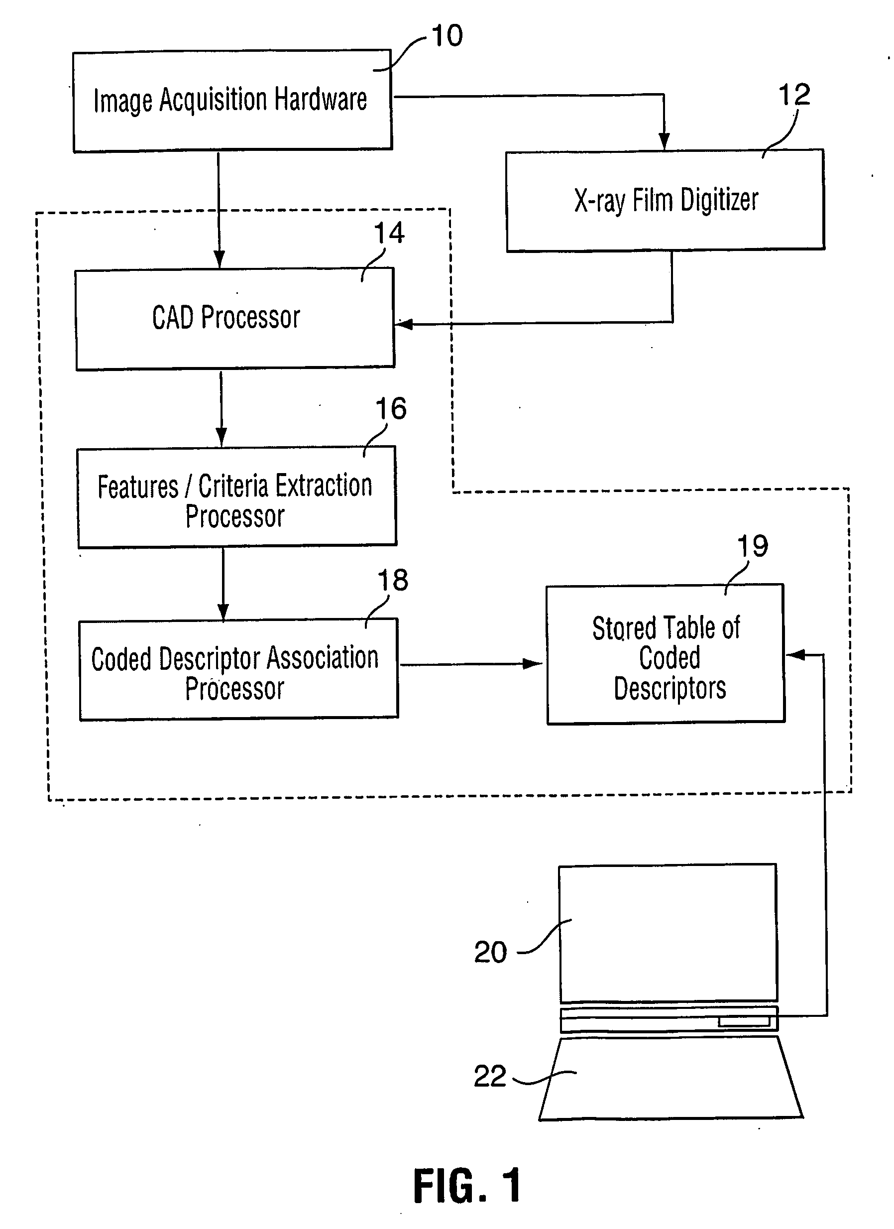
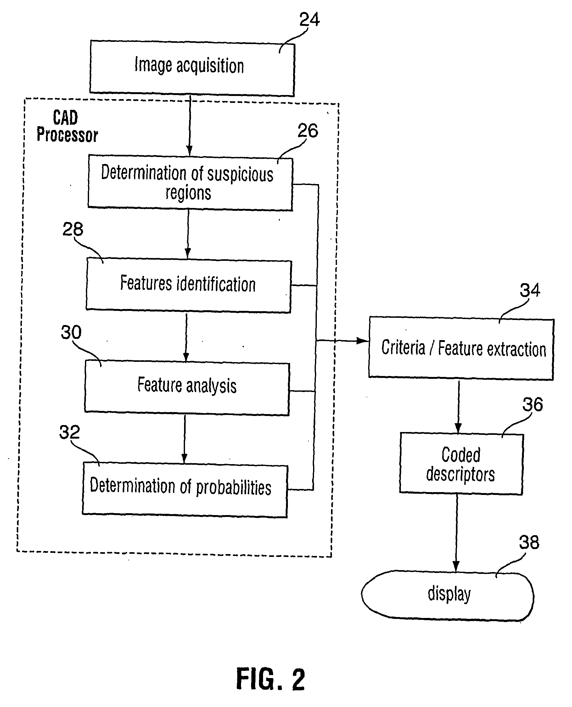
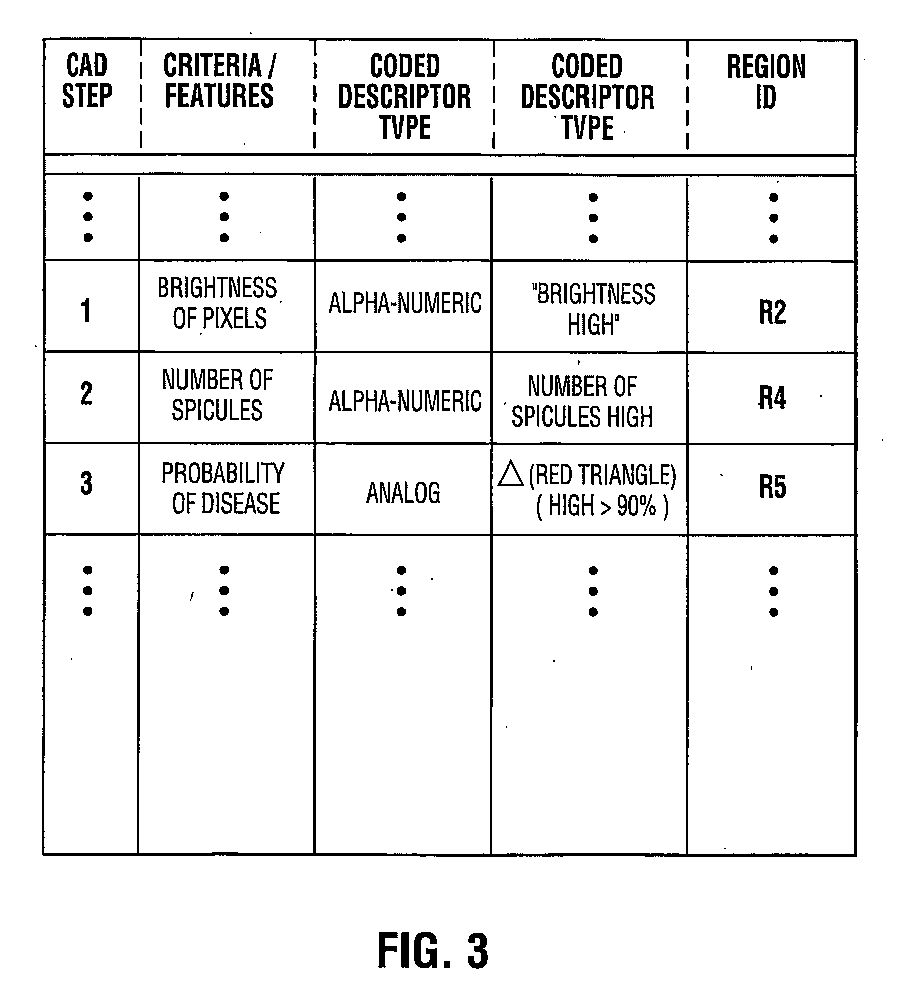
[0022] In an aspect of the present invention there is provided a system and a method for analyzing X-ray films such as mammograms using Computer Aided Detection (CAD) and for presenting the results to a user such as to indicate some or all of the criteria used by a CAD algorithm to estimate the likelihood that an abnormality in an X-ray film, indicative of a disease state in the tissue, is present. This method can assist a physician in determining the likelihood of a suspicious region being indicative of the presence of cancer or other diseases. The manner in which the results are displayed provides a simplified visual representation of the complex logic used by the computer to determine the likelihood of disease as will be further described below.

[0023] While the description of the method of the present invention will refer to analysis of mammograms for the detection of cancerous lesions, it will be appreciated that the method may also be applied to other types of diagnosis imagin...


PUM


Abstract

A method and system for Computer Aided Detection (CAD) of abnormalities in X-ray images in which the features and / or criteria used in the CAD analysis are displayed as coded descriptors on the digital image of the X-ray to provide an indication to a user of the basis underlying the identification of a particular abnormality therefore increasing the reliability of a diagnosis established by the user.

Description

[0001] This application claims priority from U.S. provisional application 60 / 413,055 filed Sep. 24, 2002 entitled “Method for Computer Aided Detection (CAD) Cued Reading of Medical Images”.TECHNICAL FIELD [0002] The present invention relates to the field of Computer-Aided Detection (CAD) of abnormalities in X-ray films. BACKGROUND OF THE INVENTION [0003] X-ray films to be analyzed by Computer Aided Detection (CAD) are scanned by a digitizer station, and converted into digital format. Computer algorithms analyze the digital images and identify features of the digitized image such as suspicious areas in the case of mammography for example. [0004] In the diagnosis process, current methods require that the physician first reads the mammograms without the aid of a computer. Once the physician has noted her findings, she turns on computer monitors that show suspicious areas identified by CAD analysis as an overlay over the digitized versions of the mammogram. The suspicious areas may conf...

Claims


Application Information

Patent Timeline
Patent Type & Authority Applications(United States)
IPC IPC(8): A61B5/00A61B6/00G06T7/00G06V20/50
CPCA61B6/502G06K9/00G06K2209/053G06T7/0012G06T2207/30068Y10S128/92G06V2201/032G06V20/50G06V2201/03G06F18/00
Inventor HARRINGTON, SCOTTMENHARDT, WIDOZHANG, DAOXIAN HEIDI
Owner CARESTREAM HEALTH INC
Why Eureka
Try Eureka