Hand held device and methods for examining a patient's retina

a technology for examining the retina of patients and hand-held devices, which is applied in the field of hand-held devices for performing retinal examination of patients' eyes, can solve the problems of preventing patients from being able to drive, work, and patients' discomfort in light, and achieves high resolution

Active Publication Date: 2006-07-06
OCUTRONICS
View PDF5 Cites 75 Cited by
  • Summary
  • Abstract
  • Description
  • Claims
  • Application Information

AI Technical Summary

Benefits of technology

[0012] In light of the above and according to one broad aspect of one embodiment of the invention, disclosed herein is a hand held device for examining a patient's retina. In one example, the device generates visible or non-visible light beams or light rays having desired spectral content that can be used, after polarization, for illuminating the patient's retina during the examination. The illuminating beams may include infrared (IR) or visible light. IR light can be used to perform the examination through the patient's undilated pupil, without the need for mydriatic drops, and the images received and processed by the hand held device can be real-time, dynamic video images of the retina. Visible light can be used for short time periods (i.e., approximately 0.1 to 0.008 seconds or other time periods) to image the retina for static or still images. Further, the hand held device may generate light selectively having two or more spectral ranges, and alternatively couple light of different spectral ranges into the optical path of the device. For instance, the illuminating beam section can provide IR light to view the retina in real-time (i.e., dynamic real time video images), then be controllably switched to provide a pulse of visible light to take a still image (i.e., a high resolution static image), then be controllably switched to again provide IR light. Hence, a hand held device according to an embodiment of the present invention can provide both video and high quality still photographic capabilities. Furthermore, in another example, the illuminating beam section can provide IR light to view the retina in real-time (i.e., dynamic real time video images), then be controllably switched to provide light with an appropriate exitation wavelength to excite fluorescein dye or other conventional dyes to perform dye studies of the retina.
[0017] According to another broad aspect of one embodiment of the present invention, disclosed herein is a hand held device for examining a retina through a pupil. In one example, the hand held device may include a light source generating light (visible or non-visible); a first lens refracting the light to form a refracted beam; a first polarizer receiving the refracted beam and forming a linear polarized beam; a second polarizer for reflecting linearly polarized light, the second polarizer receiving the linear polarized beam and reflecting the linear polarized beam; and a second lens refracting the linear polarized beam from the second polarizer, the second lens converging the linear polarized beam for passage through the pupil to illuminate the retina. This illumination of the retina can produce a retinal image that is received and processed by the hand held device. Further, the device can be used to examine the patient's retina through an undilated pupil, without the need for mydriatic drops.
[0019] The light source may include two or more discrete light sources providing different spectral ranges of illumination, for example, a first lamp in series with an infrared (IR) bandpass filter to generate IR light, a second lamp in series with an visible light bandpass filter to generate visible light, wherein when the first lamp is on, the second lamp is off. A simple electronic switch may be used to provide power to the first lamp and not the second, and then to provide power to the second lamp and not the first. In this manner, the device can switch between two different spectral ranges of illumination, without the need for moving parts.

Problems solved by technology

Once a conventional eye exam is completed, the patient's pupils remain dilated for possibly hours, which may be very inconvenient for the patient and may prevent the patient from being able to drive, work, etc.
Even with the use of filters, the light is uncomfortable to the patient, sometimes to the point of making the examination difficult or impossible.
Opacities to visible light located in the lens of the eye (e.g., cataract) or in the vitreous (e.g., blood) also limit the ability to see the retina and optic nerve, sometimes totally precluding such examination.
Some smaller variants have been developed, but still require cables hooked to large power supplies, resulting in cumbersome units.
The large camera units generally may require the services of a professional photographer to operate, and the units may be priced in the range from $35,000 to $100,000, both of which factors limit their availability.
The table mountings can make them inaccessible to wheel-chair patients, unless special table extensions are available.
The cameras are rarely available for bed patients, in some instances.
Also, such cameras are difficult to move from office to office, except by van.

Method used


Image

  • Hand held device and methods for examining a patient's retina
  • Hand held device and methods for examining a patient's retina
  • Hand held device and methods for examining a patient's retina

Examples


Embodiment Construction

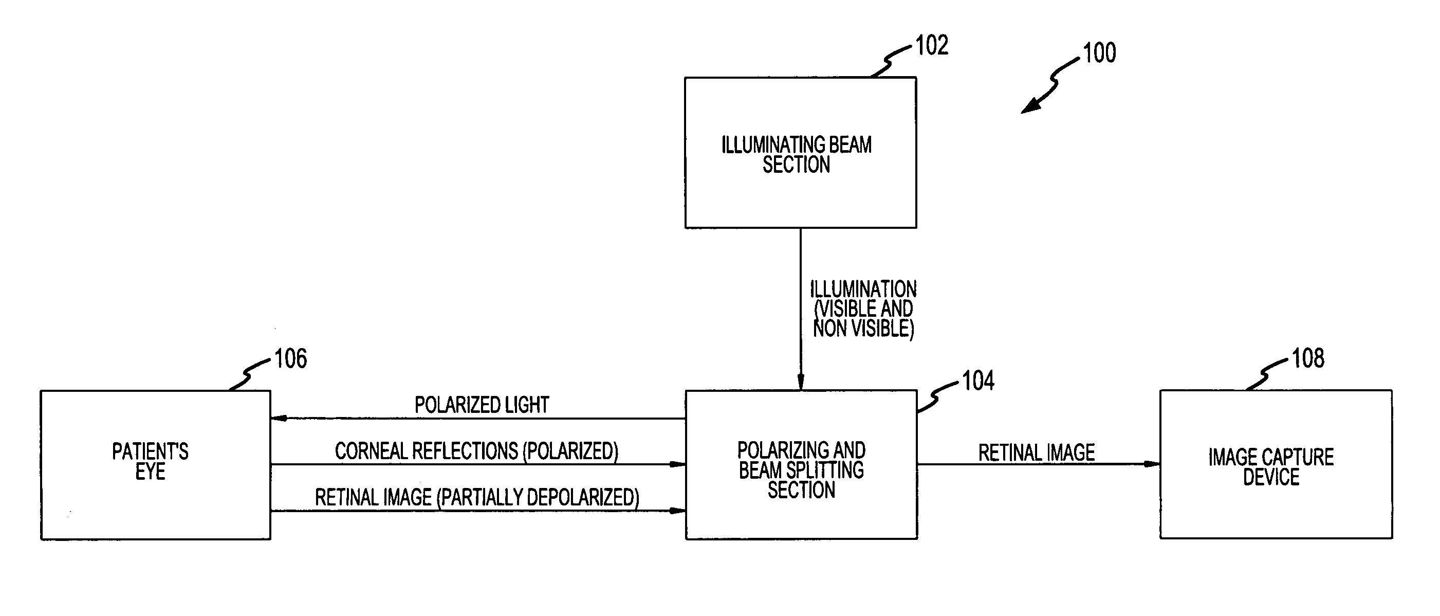
[0065] According to one embodiment of the present invention, disclosed herein is a hand held device for examination of a patient's retina. Embodiments of the present invention permit a doctor to examine the retina of a patient without the use of mydriatic eye drops and therefore without dilating the pupils. As will be described below, some embodiments of the present invention use a combination of infrared (IR) and visible light, along with various optical techniques, to capture images (such as static still images and dynamic video images) of a patient's retina. Embodiments of the device and methods may be used to examine patient's eyes, such as the retina of a human or an animal.

[0066]FIG. 1 illustrates a block diagram of an example of hand held device for examining a patient's retina, in accordance with one embodiment of the present invention. An illuminating beam section generates visible or non-visible light beams or light rays having desired spectral content that can be used, a...


PUM


Abstract

A hand held device for examining a patient's retina. In one example, the device generates visible or non-visible illuminating beams having desired spectral content. The illuminating beams are then polarized to generate polarized light beam(s) that are directed towards the patient's retina. At the patient's eye, a portion of the polarized beam is partially reflected by the cornea and travels back to the device, and another portion of the polarized beam enters the eye through the undilated pupil, illuminates the patient's retina, and generates a reflected retinal image (generally de-polarized) which also travels back to the device. The device receives the retinal image and transmits certain portions of the retinal image on through to an image detector (i.e., CCD or CMOS); and the device deflects the received polarized corneal reflections away from the image capture device. Hence, the image capture device receives retinal images but does not receive the polarized corneal reflected light.

Description

CROSS-REFERENCE TO RELATED APPLICATION [0001] This application claims priority under 35 U.S.C. 119(e) to U.S. Provisional patent application No. 60 / 542,639 entitled “HAND HELD DEVICE AND METHODS FOR EXAMINING A PATIENT'S RETINA” filed Feb. 5, 2004, the disclosure of which is hereby incorporated by reference in its entirety.FIELD OF THE INVENTION [0002] This invention relates, in general, to methods and devices for performing an examination of the eye, and more particularly, for performing an examination of the retina of a patient's eye. BACKGROUND OF THE INVENTION [0003] Many conventional retinal eye examinations involve the use of eye drops which are given to a patient to force the patient's iris open (dilates the pupils). These drops are known as mydriatic eye drops. After the patient's pupils are dilated, the eye doctor then examines the patient's retina, also known as the “fundus,” with a conventional ophthalmoscope, which typically uses white light. Once a conventional eye exam...

Claims


Application Information

Patent Timeline
Patent Type & Authority Applications(United States)
IPC IPC(8): A61B3/10
CPCA61B3/1208
Inventor COLLINS, WILLIAM J.LEVINE, ROBERT E.
Owner OCUTRONICS
Why Eureka
Try Eureka