MRI Biopsy Device

a biopsy device and tissue technology, applied in medical science, surgery, vaccination/ovulation diagnostics, etc., can solve the problem that the clinician may find the control module inconveniently remote, and achieve the effect of facilitating user control

Inactive Publication Date: 2006-11-16
DEVICOR MEDICAL PROD
View PDF100 Cites 151 Cited by
  • Summary
  • Abstract
  • Description
  • Claims
  • Application Information

AI Technical Summary

Benefits of technology

[0010] The invention overcomes the above-noted and other deficiencies of the prior art by providing a handpiece of a magnetic resonance imaging (MRI) compatible core biopsy system that includes a graphical user interface that facilitates user control even with vacuum, power generation, and control

Problems solved by technology

While a full function GUI has numerous clinical benefits, the clinician may find t

Method used


Image

  • MRI Biopsy Device
  • MRI Biopsy Device
  • MRI Biopsy Device

Examples


Embodiment Construction

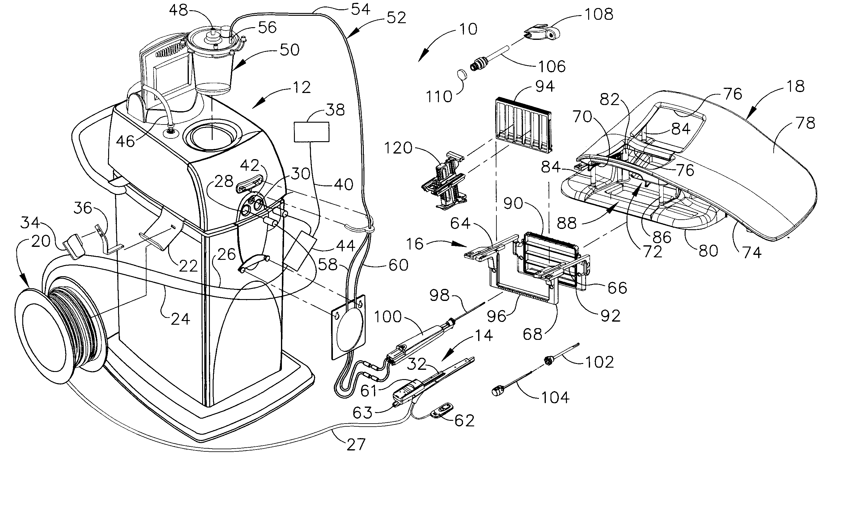
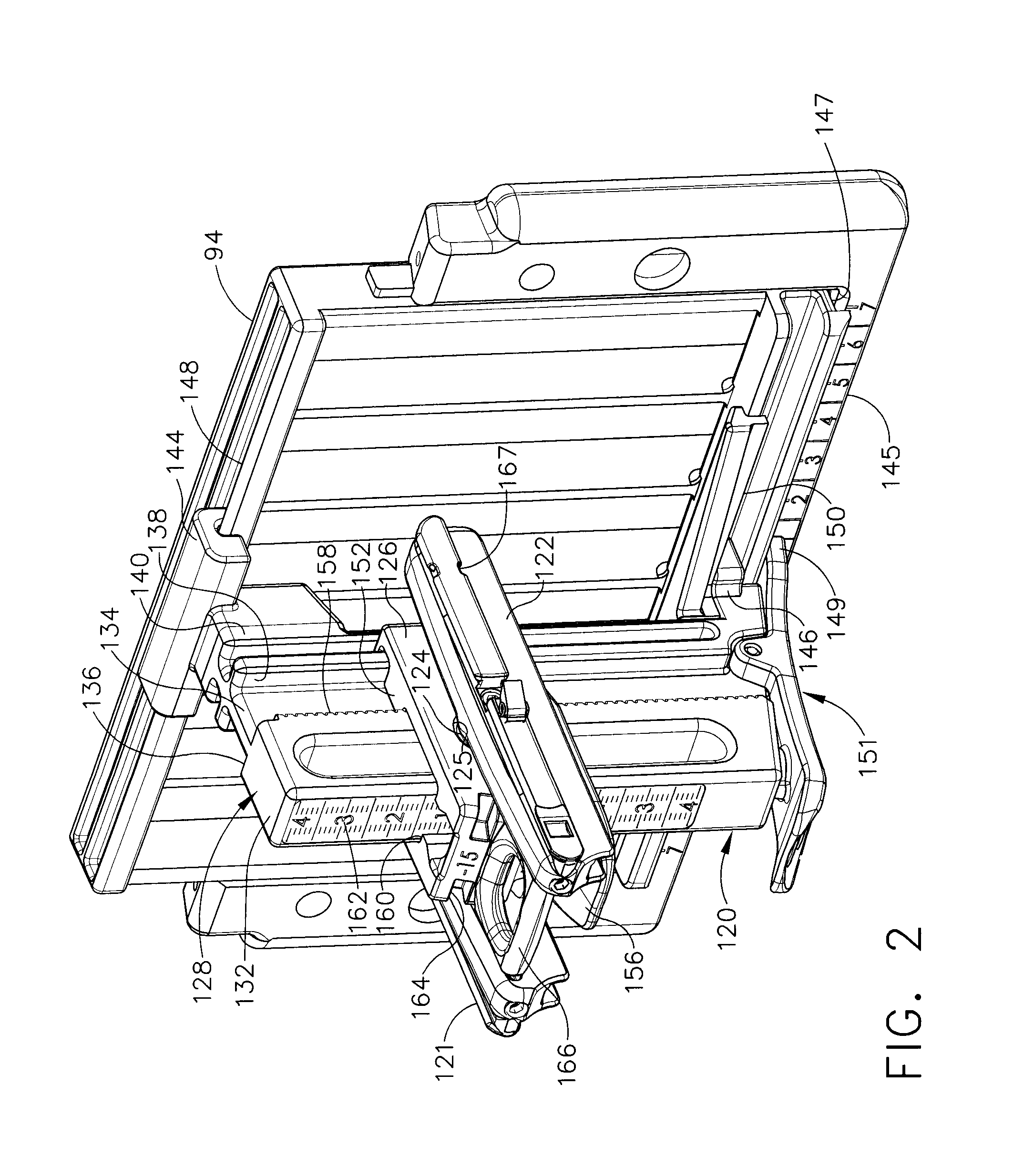
[0027] An MRI biopsy device advantageously includes is partially disposable for sterility purposes with a reusable portion for economy. Inconvenience of mechanical, electrical, and pneumatical coupling to a remotely placed control portion, necessitated by a strong magnetic field and sensitive RF receiving components of an MRI machine, is mitigated. First, proximal detachable intuitive controls and displays on the MRI biopsy device give interactive control even after insertion into localizing and guiding structures. Second, binding of mechanical coupling to the MRI biopsy device is sensed prior to equipment damage or malfunction. Third, mechanical coupling is moved closer to engagement points between the MRI biopsy device and guiding structures to reduce torque loads, especially those transferred through its distal probe. Fourth, a single mechanical drive cable drives a fixed ratio transmission that translates and rotates a cutter of the distal probe to realize an effective fixed rat...


PUM


Abstract

A magnetic resonance imaging (MRI) compatible core biopsy system uses a biopsy device having intuitive graphical displays and a detachable remote keypad that advantageously allows convenient control even within the close confines afforded by a localization fixture installed within a breast coil that localizes a patient's breast and guides a probe of the biopsy device relative to the localized breast. A control module for interactive control and power generation are remotely positioned and communicate and transmit rotational mechanical energy via sheathed cable.

Description

CROSS REFERENCE TO RELATED APPLICATIONS [0001] The present application is a continuation in part of U.S. patent application Ser. No. 11 / 103,959, “MRI BIOPSY DEVICE LOCALIZATION FIXTURE” to Hughes et al., filed on 12 Apr. 2005, the disclosure of which is hereby incorporated by reference in its entirety.FIELD OF THE INVENTION [0002] The present invention relates, in general, to a method of imaging assisted tissue sampling and, more particularly, to an improved method for positioning a biopsy probe with respect to a magnetic resonance imaging (MRI) breast coil for acquiring subcutaneous biopsies and for removing lesions. BACKGROUND OF THE INVENTION [0003] Core biopsy devices have been combined with imaging technology to better target a lesion in breast tissue. One such commercially available product is marketed under the trademark name MAMMOTOME™, by Ethicon Endo-Surgery, Inc. An embodiment of such a device is described in U.S. Pat. No. 5,526,822 issued to Burbank, et al., on Jun. 18, ...

Claims


Application Information

Patent Timeline
IPC IPC(8): A61B10/00
CPCA61B10/0041A61B2019/5487A61B10/0283A61B17/3403A61B19/201A61B19/52A61B2017/00128A61B2017/00199A61B2017/00212A61B2017/00796A61B2017/00911A61B2017/3411A61B2018/00928A61B2019/205A61B2019/4857A61B2019/5236A61B2019/5408A61B2019/5454A61B10/0275A61B90/17A61B90/36A61B90/11A61B2090/0811A61B2090/374A61B2090/3908A61B2090/3954A61B2090/3987A61B10/0266A61B2010/0208A61B5/708A61B2560/0266A61B2560/0462A61B5/065A61B5/743A61B5/7435
Inventor HABERSTICH, WELLS D.RAGHAVENDRAN, RAJ G.HIBNER, JOHN A.
Owner DEVICOR MEDICAL PROD
Why Eureka
Try Eureka