Method for extending the display of a multi-dimensional image of an object region

a multi-dimensional image and object technology, applied in image enhancement, angiography, instruments, etc., can solve the problem of only offering a limited field of view for image captur

Active Publication Date: 2006-11-23
SIEMENS HEALTHCARE GMBH
View PDF6 Cites 18 Cited by
  • Summary
  • Abstract
  • Description
  • Claims
  • Application Information

AI Technical Summary

Benefits of technology

[0006] An object of the present invention is to provide a method for extension of the display range of 2D image recordings of an object area which offers advantages precisely when using mobile compact devices for the capture of 2D image recordings.
[0009] In a preferred embodiment of the inventive method, the additional 2D image data are therefore also acquired with a higher resolution than the first 2D or 3D image data. The image display of the larger object area thus is adapted to the higher resolution of the integrated display of the additional 2D image data by interpolation. In another embodiment, the additional 2D image data are also acquired with a higher image contrast or with different characteristics (e.g. by using tracers or contrast substances) than the first 2D or 3D image data. This leads to a locally better and more current detail display in a limited area of the combined image display. The synthesis of the two images is performed based on a correct location and orientation indexing, as well as preferably with a calibration during the 2D recording.
[0012] For instance, in a first variant at least three spatially uniquely identifiable marks can be attached to the patient that remain on the patient for both the first earlier recording of the 3D image data set as well as for the later additional 2D image data. To increase accuracy and reliability of identification of each mark, more than three marks can be used. The marks can be used for image registration of the current patient position with respect to the earlier recorded first 3D data set. Moreover, from the position detectable in the 2D image data, the image perspective relative to the first 3D image data set is determined with which the additional 2D image data was obtained. Knowledge of this image (data acquisition) perspective or projection geometry of the additional 2D image data enables the calculation, in combination with the registration of the first 3D image data set, of an image display of the larger image area through superimposition of the different irradiated layers according to this perspective, so that a correct combination of the two images is possible.

Problems solved by technology

These modalities, however, offer only a limited field of view for image capture.

Method used


Image

  • Method for extending the display of a multi-dimensional image of an object region
  • Method for extending the display of a multi-dimensional image of an object region
  • Method for extending the display of a multi-dimensional image of an object region

Examples


Embodiment Construction

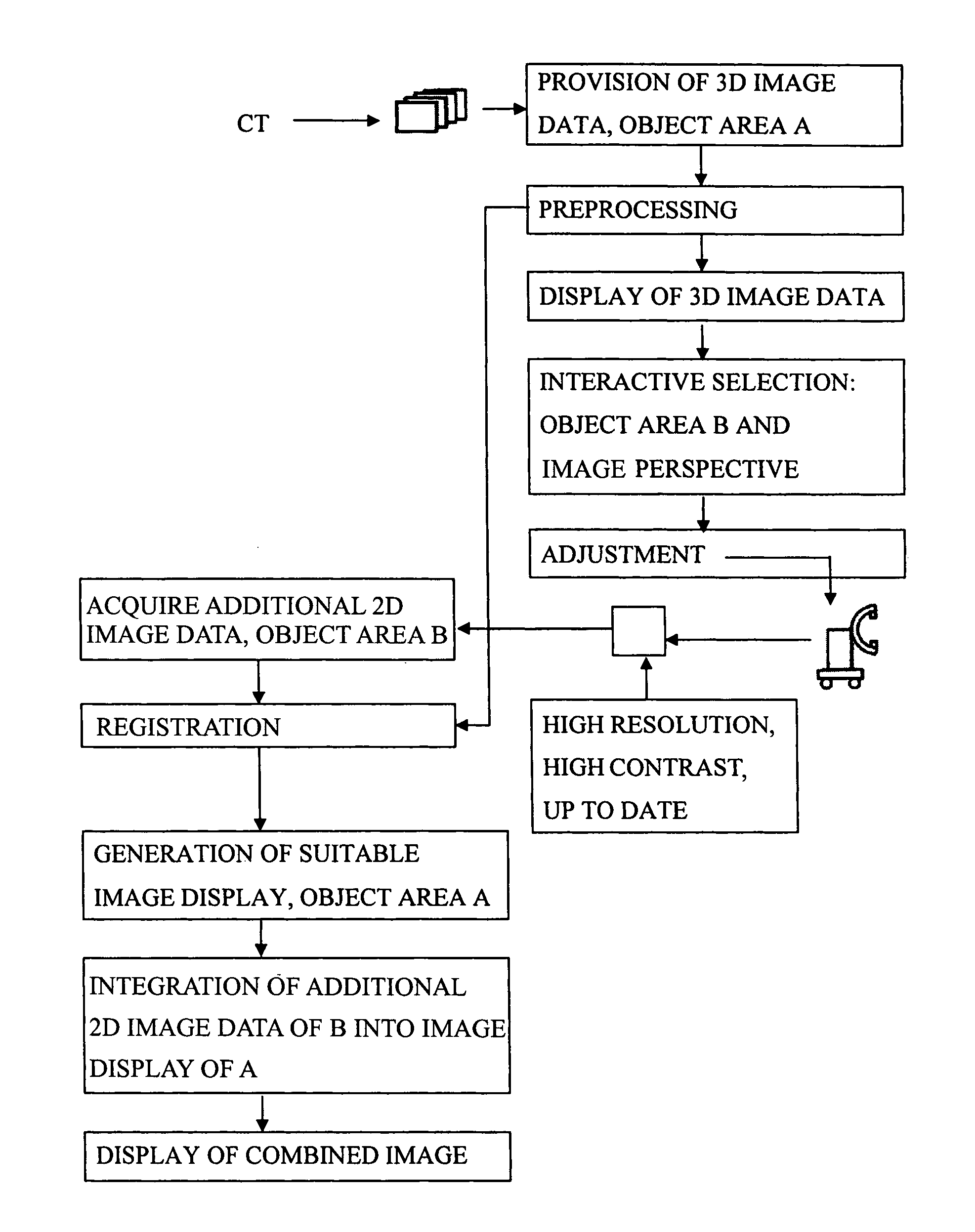
[0024] The inventive method is explained in detail using the example of an operation, for instance after a pelvic fracture, in which during the operation additional 2D X-ray data of the treatment area are obtained using a mobile C-arm device and displayed.

[0025] Both for the planning and for the performance of an operation nowadays, a series of first 3D image data sets are used which are obtained before the intervention, for example from CT volume recordings. Through visualization of this first 3D image data, a complete, large-scale overview is obtained of the entire relevant body environment. In an operation, the physician must rely on a current image exposure for different reasons, which is executed for instance using endoscopy, X-ray illumination, or with radiographical individual recordings. In the case of repeated additional image exposures during the operation, the physician can track changes immediately in this manner. The current image exposure is already required for safet...


PUM


Abstract

In a method to extend the display range of 2D image recordings of an object region, particularly in medical applications, first 2D or 3D image data are acquired from a larger object region, and at least one additional set for 2D image data of a smaller object region is acquired that lies within the larger object region The first 2D or 3D image data are brought into registration with the additional 2D image data with a projection geometry. From the first 2D or 3D image data, an image data set is generated for an image display of the first object region, which is suitable for combination with the additional 2D image data. In the image display of the larger object region, at least temporarily, at least one display of the additional 2D image data is integrated, by image data in the first image data set, for the image display of the larger object region, being replaced with image data from the additional 2D image data representing the smaller image region. An overview of the larger object region is thus enabled, with the smaller object region of interest being displayed within the image in a more up-to-date fashion, as well as with higher resolution and / or higher contrast.

Description

BACKGROUND OF THE INVENTION [0001] 1. Field of the Invention [0002] The present invention concerns a method for the extension of the display range of 2D images of an object region of the type wherein first 2D or 3D image data of a larger object region are provided, at least one set of additional 2D image data of a smaller object region is obtained that lies within the larger object region, and the additional image data are displayed in combination with the 2D image. [0003] 2. Description of the Prior Art [0004] In surgical interventions nowadays, compact image capture systems such as mobile X-ray devices, ultrasound devices, or endoscopes / laparoscopes are often used. These modalities, however, offer only a limited field of view for image capture. For the surgeon, however, it is desirable to be able to view the local situation in the treatment area in a larger context as well. [0005] Different options are known in which 3D image data of the body are recorded at some larger time previ...

Claims


Application Information

Patent Timeline
Patent Type & Authority Applications(United States)
IPC IPC(8): G06T11/20A61B1/00A61B1/04A61B5/00A61B6/00A61B6/03A61B8/00G09G5/00G09G5/14G09G5/36H04N7/18
CPCA61B6/12A61B6/4405A61B6/4441A61B6/466A61B6/504G06T2207/30004A61B8/483A61B8/5238A61B2019/5289G06T7/0024G06T19/00A61B6/5235A61B2090/364G06T7/30
Inventor BARFUSS, HELMUTBARTH, KARLWESSELS, GERD
Owner SIEMENS HEALTHCARE GMBH
Why Eureka
Try Eureka