Extraocular muscle prosthesis

a prosthesis and extraocular muscle technology, applied in the field of extraocular muscle prosthesis, can solve the problems of reducing visual acuity and anterior segment ischemia, and achieve the effect of restoring eyelid function

Inactive Publication Date: 2006-12-21
UNIV OF MIAMI
View PDF19 Cites 11 Cited by
  • Summary
  • Abstract
  • Description
  • Claims
  • Application Information

AI Technical Summary

Benefits of technology

[0008] The invention may also be embodied in a prosthetic device for restoring eyelid function comprising: a housing; a coil spring component disposed in said housing; a proximal connector operatively connected to a proximal end of the biasing component; and a distal connector operatively connected to a distal end of the biasing component, wherein said proximal connector is configured for being secured with respect to an orbital bone and said distal connector adapted to be secured to the upper eyelid.

Problems solved by technology

Unfortunately, operating on the remaining three rectus muscles could compromise the blood supply to the eye, leading to anterior segment ischemia and reduced visual acuity.
All these surgical options share one major drawback—the globe is tethered to one position, unable to adduct or abduct the eye.

Method used


Image

  • Extraocular muscle prosthesis
  • Extraocular muscle prosthesis
  • Extraocular muscle prosthesis

Examples


Embodiment Construction

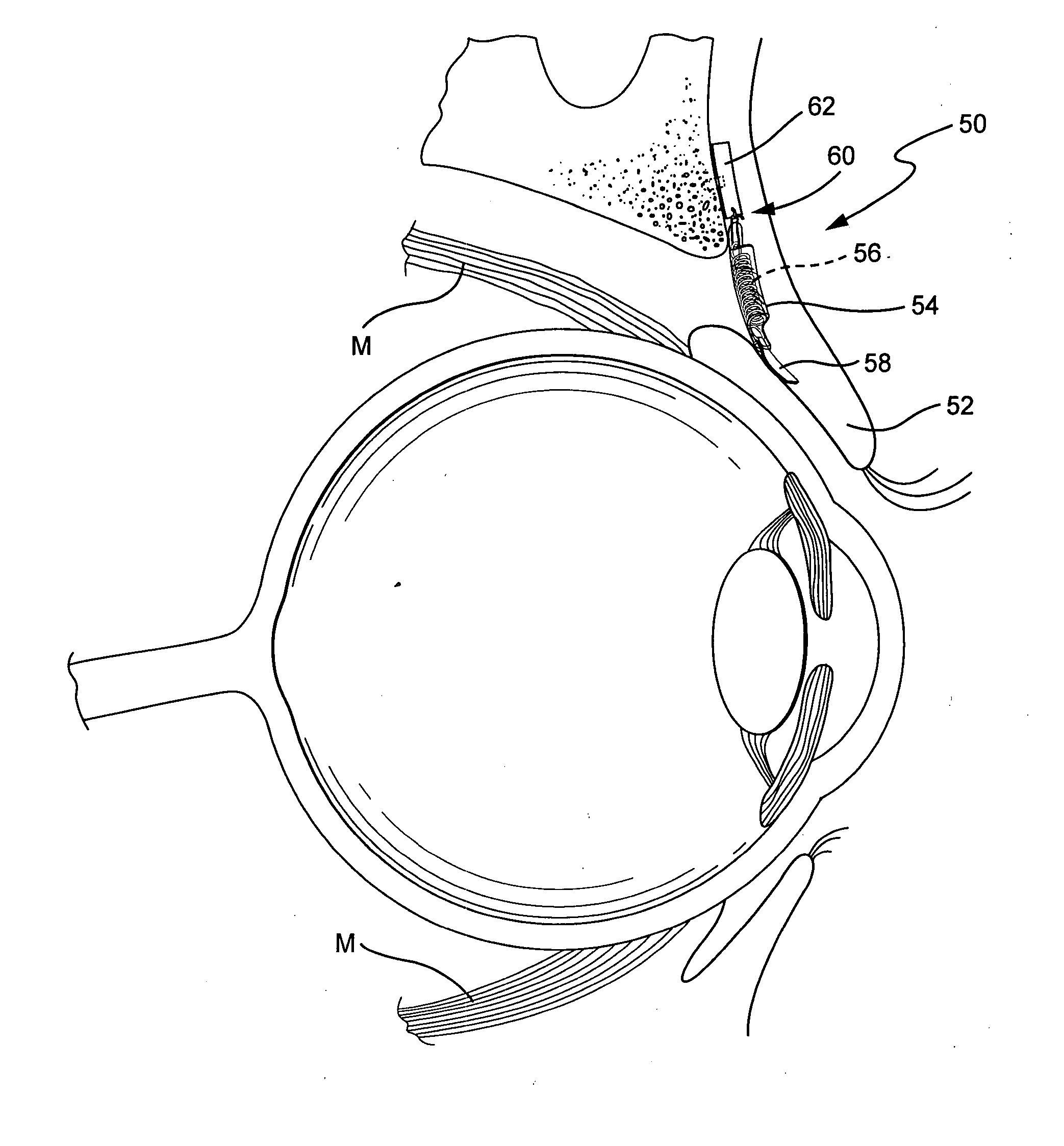
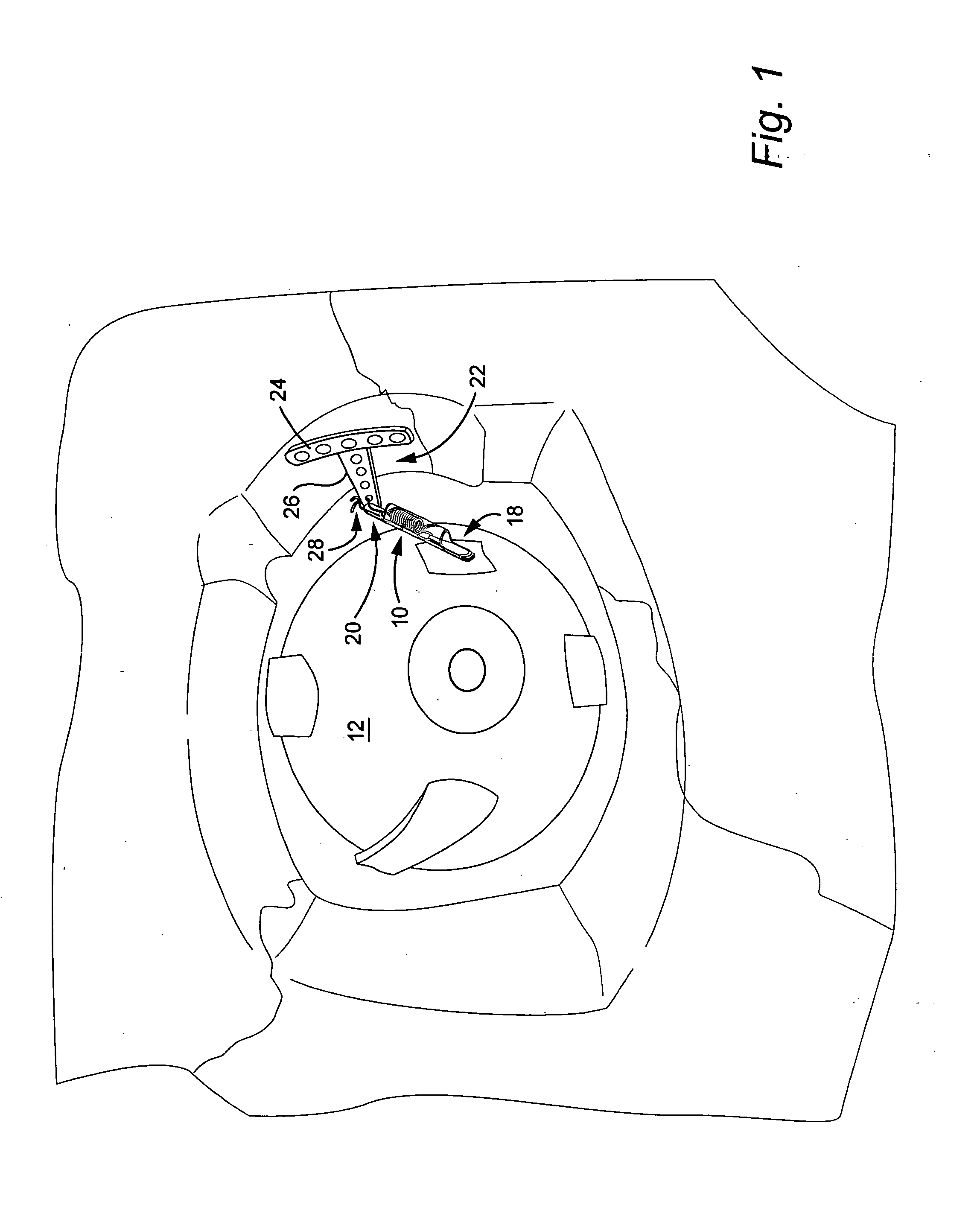
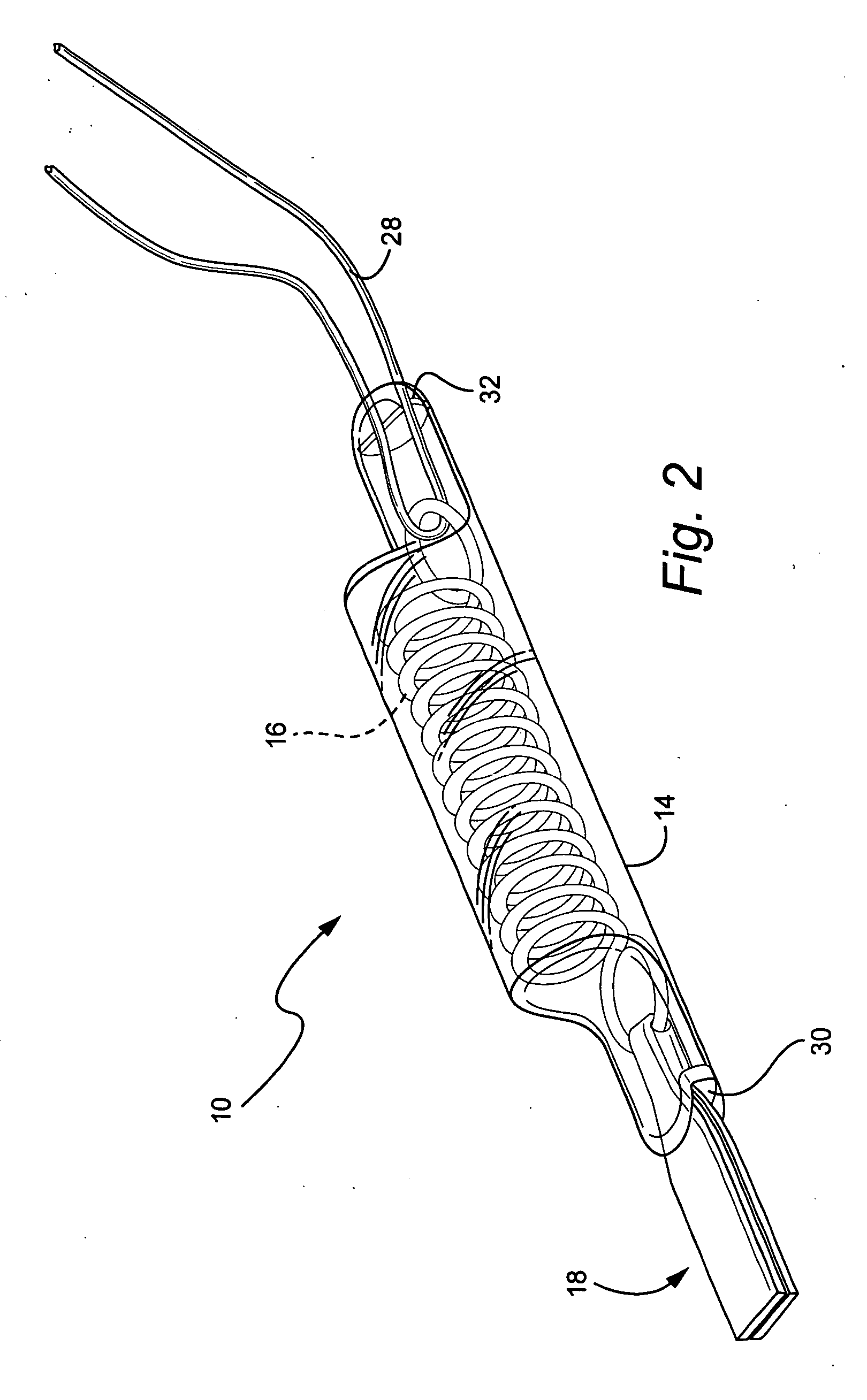
[0013] An eye muscle prosthesis is provided in an example embodiment of the invention to replace a paralyzed or absent extraocular muscle. More specifically, an eye muscle prosthesis is provided comprising a spring device 10 for biasing the globe 12 of the eye to the primary position. The spring device includes an outer housing 14, a component 16 for biasing the globe to the primary position, a connector 18 for operatively connecting one end of the biasing component to the globe, and a connector 20 for operatively connecting the other end of the biasing component to the boney perimeter of the orbit.

[0014] In the illustrated example embodiment, the biasing component is a coil torsion spring. In an example embodiment of the invention, the spring is formed from Elgiloy®, although other materials such as titanium, Nitenol, stainless steel or the like, which are known for their biocompatibility and resistance to corrosion as well as suitability for minute spring components, could be use...


PUM


Abstract

A prosthetic device and method to restore extraocular muscle function. The device includes a housing; a biasing component disposed in the housing; a proximal connector operatively connected to a proximal end of the biasing component; and a distal connector operatively connected to a distal end of the biasing component, wherein the proximal connector is configured for being secured with respect to an orbital bone and the distal connector adapted to be secured to the paralyzed or absent muscle stump, e.g. on the globe, or to the eyelid.

Description

CROSS-REFERENCE TO RELATED APPLICATION [0001] This application claims the benefit of Provisional Application No. 60 / 690,897, filed Jun. 16, 2005, the entire content of which is hereby incorporated by reference in this application.BACKGROUND OF THE INVENTION [0002] Classic examples of paralyzed extraocular muscle are congenital or acquired complete third nerve palsy and ocular fibrosis syndrome. An ophthalmic complication encountered with increasing frequency in functional endoscopic sinus surgery (FESS) is inadvertent amputation of the medial rectus muscle. The consequence of such medial rectus dysfunction is that the involved eye assumes a large angle exotropia, an inability to adduct the eye, and disabling diplopia. Occasionally, severe paralytic and restrictive or absent extraocular muscle will require globe fixation in the primary position because of the presence of an active antagonist muscle. [0003] Surgical remedies include supramaximal recession of the lateral rectus and tra...

Claims


Application Information

Patent Timeline
Patent Type & Authority Applications(United States)
IPC IPC(8): A61F2/14A61F2/08
CPCA61B17/8061A61F2002/0894A61F9/0017A61F2/08
Inventor TSE, DAVID T.
Owner UNIV OF MIAMI
Why Eureka
Try Eureka