Image processing apparatus and X-ray CT apparatus

Inactive Publication Date: 2007-01-25
GE MEDICAL SYST GLOBAL TECH CO LLC
View PDF16 Cites 21 Cited by
  • Summary
  • Abstract
  • Description
  • Claims
  • Application Information

AI Technical Summary

Benefits of technology

[0008] Therefore, an object of the present invention is to provide an image processing apparatus that uses pieces of information on the direction of a time axis and the directions of spatial axes to improve the quality of a four-dimensional image that is a time-varying three-dimensional image and that is composed of time-sequential three-dimensional images, a three-dimensional image that is a time-varying two-dimensional image and that is composed of time-sequential two-dimensional image, or an N-dimensional image that is a time-varying N-1-dimensional image and that is composed of time-sequential N-1-dimensional images.
[0009] Another object of the present invention is to provide an X-ray CT apparatus that includes a matrix-type two-dimensional area X-ray detector represented by a multi-array X-ray detector or a flat-panel X-ray detector, that offers target image quality with a smaller X-ray dose by improving the quality of time-sequential three-dimensional images or time-sequential two-dimensional images, which are produced through conventional (axial) scanning, cine scanning, or helical scanning, using pieces of information on the direction of a time axis and the directions of spatial axes.
[0010] The present invention provides an image processing method and an image processing apparatus that can improve the quality of a four-dimensional image that is a time-varying three-dimensional image, a three-dimensional image that is a time-varying two-dimensional image, or an N-dimensional image that is a time-varying N-1-dimensional image (an N-1-dimensional image defined with N-1 independent parameters as a base) by performing spatial filtering or adaptive spatial filtering in the direction of a time axis and the directions of spatial axes.

Problems solved by technology

From this viewpoint, the related art is not fully acceptable.
Moreover, in the field of medical-purpose radiographic diagnosis, the issue of an unnecessary patient X-ray dose has become more and more controversial along with prevalence of X-ray CT apparatuses and diversity in purposes of examination.

Method used


Image

  • Image processing apparatus and X-ray CT apparatus
  • Image processing apparatus and X-ray CT apparatus
  • Image processing apparatus and X-ray CT apparatus

Examples


Embodiment Construction

[0078] The present invention will be described by taking an illustrated embodiment for instance. Noted is that the present invention will not be limited to the embodiment.

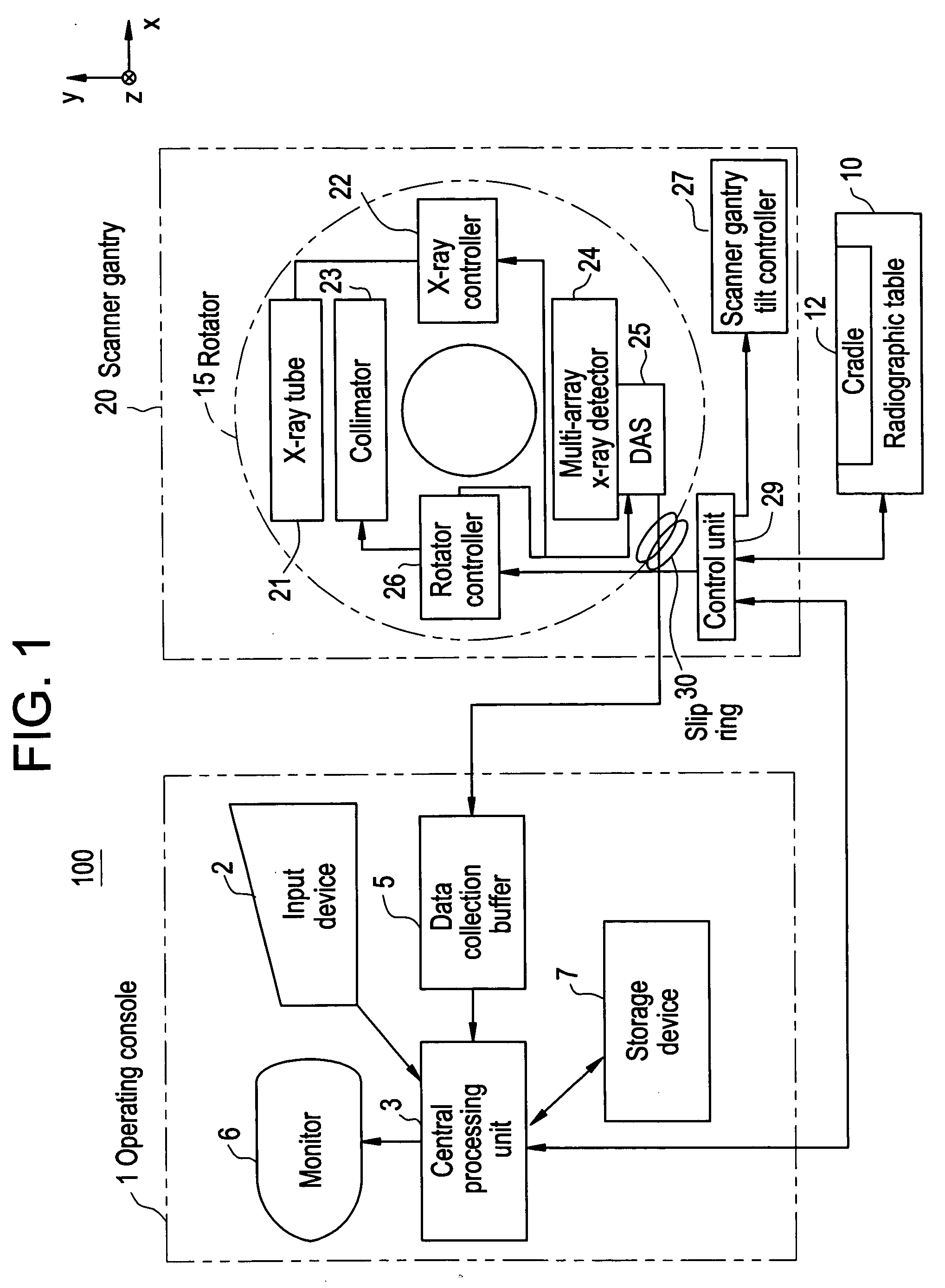
[0079]FIG. 1 is a block diagram showing the configuration of an X-ray CT apparatus in accordance with an embodiment of the present invention. The X-ray CT apparatus 100 includes an operator console 1, a radiographic table 10, and a scanner gantry 20.

[0080] The operator console 1 includes an input device 2 that receives an operator's entry, a central processing unit 3 that performs preprocessing, image reconstruction, post-processing, and others, a data collection buffer 5 in which X-ray detector data items acquired by the scanner gantry 20 are collected, a monitor 6 on which a reconstructed tomographic image is displayed according to projection data items produced by performing preprocessing on the X-ray detector data items, and a storage device 7 in which programs, X-ray detector data items, projection data item...


PUM


Abstract

The present invention is intended to improve the quality of a four-dimensional image that is a time-varying three-dimensional image. The four-dimensional image that is a time-varying three-dimensional image is spatially filtered in the direction of a time axis and the directions of spatial axes alike.

Description

CROSS REFERENCE TO RELATED APPLICATIONS [0001] This application claims benefit of Japanese Application No. 2005-210334 filed Jul. 20, 2005. BACKGROUND OF THE INVENTION [0002] The present invention relates to an image processing apparatus that improves the quality of a time-varying image, or more particularly, to improvement in the quality of images produced through helical scanning or cine scanning performed by an X-ray CT apparatus, and reduction in a patient dose derived from the helical scanning or cine scanning. [0003] In an X-ray CT apparatus including a two-dimensional X-ray area detector represented by a multi-array X-ray detector or a flat-panel X-ray detector, a three-dimensional spatial filter defined in the directions of x, y, and z spatial axes as shown in FIG. 12(b) is applied to a three-dimensional image produced through cine scanning and defined in the directions of the x, y, and z axes as shown in FIG. 12(a). Noises are thus minimized. An example of the three-dimensi...

Claims


Application Information

Patent Timeline
IPC IPC(8): G06K9/00
CPCG06T11/005A61B6/027G06T2211/412A61B6/03G01T1/00A61B6/00
Inventor NISHIDE, AKIHIKOHAGIWARA, AKIRAHORIUCHI, TETSUYA
Owner GE MEDICAL SYST GLOBAL TECH CO LLC
Why Eureka
Try Eureka