
Diagnostic markers of wound infection III

a technology of diagnostic markers and wound infection, applied in the field of monitoring patients, can solve the problems of slow healing, further necrosis of wounds, and difficult to achieve the mass of specimens required for invasive procedures

Inactive Publication Date: 2007-03-08
WOUNDCHECK LAB US
View PDF1 Cites 4 Cited by
  • Summary
  • Abstract
  • Description
  • Claims
  • Application Information

AI Technical Summary

Benefits of technology

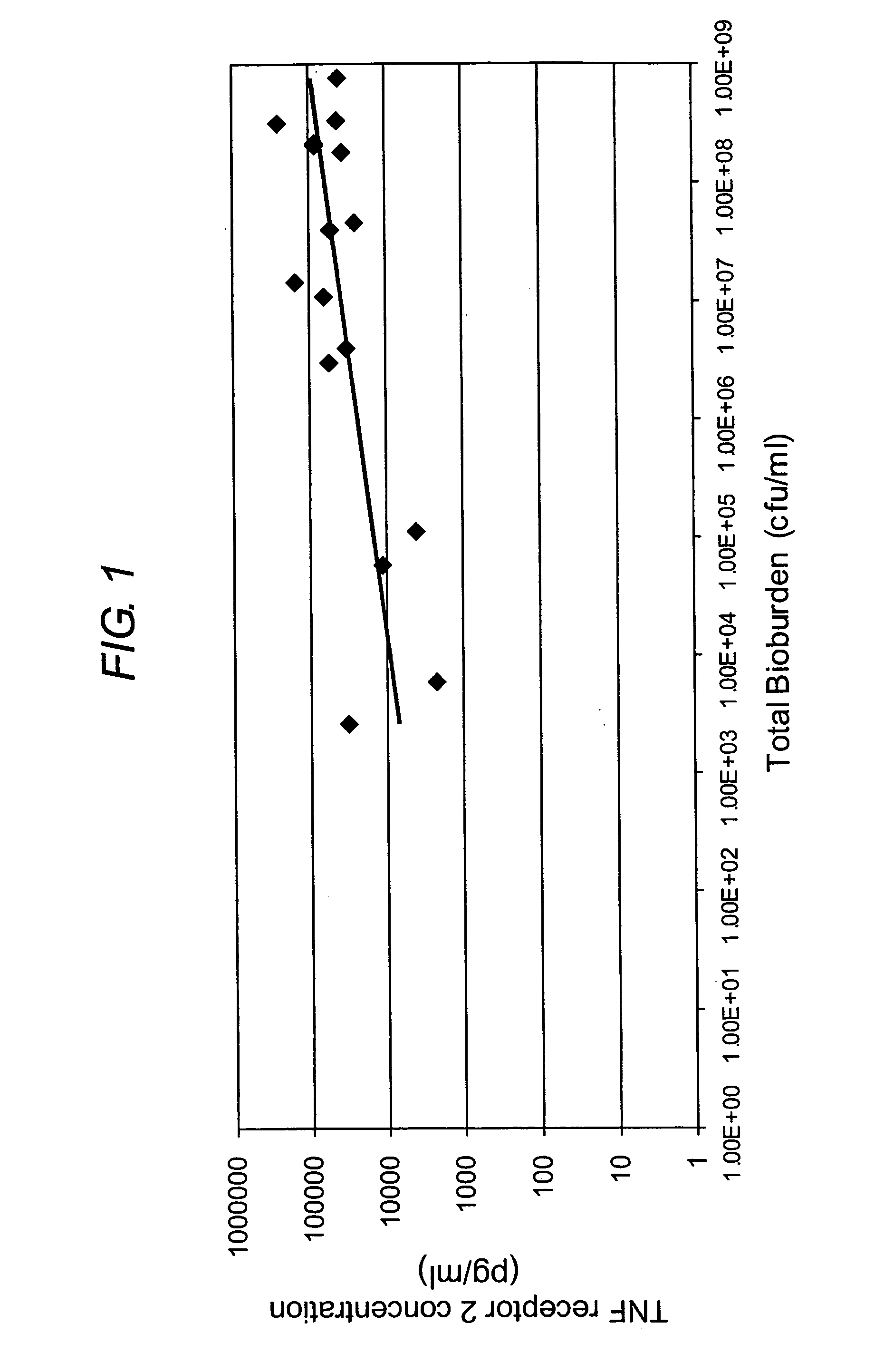
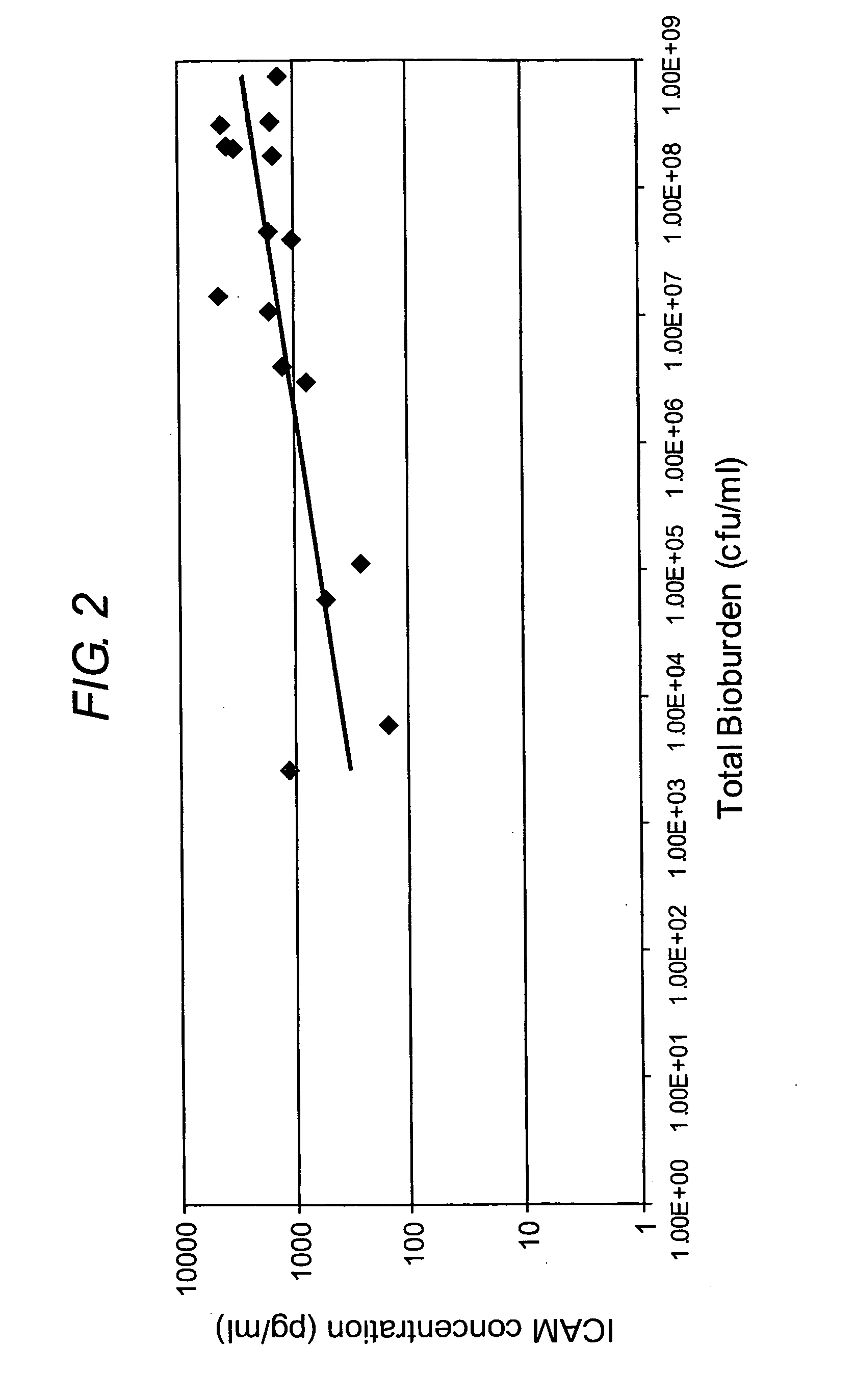
[0007] The present inventors have made the surprising discovery that elevated levels of cell surface receptors, in particular ICAM 1 and TNF-RII, are indicative of wound infection, and that the level of the said cell surface receptors in wound fluid is correlated to the level of bioburden in the woun...

Problems solved by technology

Lastly, infection can result in a prolonged inflammatory phase and thus slow healing, or may cause further necrosis of the wound.
Tissue biopsy provides the most accurate results, but this is an invasive procedure that is difficult to achieve for the mass of specimens required.
Furthermore, microbiological diagnosis of wound infection can take 48 to 72 hours, which allows time for infection to further develop if first-line / best-guess treatment is not employed immediately.

Method used


Image

  • Diagnostic markers of wound infection III
  • Diagnostic markers of wound infection III

Examples


example 1

Collection and Treatment of Wound Fluid—Removal of Infected and Non-Infected Wound Fluid

[0075] All patients enrolled in the study had venous leg ulcers of at least 30 days duration and a surface area of at least 1 cm2. Patients were diagnosed as ‘non-infected, normal appearance of wound, or ‘infected’ based on a minimum of 4 clinical signs and symptoms indicative of infection. Patients were excluded from the study if exposed bone with positive osteomyelitis was observed. Other exclusion criteria included concomitant conditions or treatments that may have interfered with wound healing and a history of non-compliance that would make it unlikely that a patient would complete the study. Wound fluids were collected from the patients following informed consent being given from all patients or their authorized representatives. The protocol was approved by the Ethics Committee at the participating study center prior to commencement of the study. The study was conducted in accordance with ...


PUM

PropertyMeasurementUnit
Timeaaaaaaaaaa
Timeaaaaaaaaaa
Surfaceaaaaaaaaaa

Abstract

The present invention relates to a method of diagnosis or prognosis of a mammalian wound infection, said method comprising the step of measuring the level of at least one cell surface receptor in a sample of wound fluid. The preferred cell surface receptors are Intercellular adhesion molecule-1 (ICAM 1) and Tumor Necrosis Factor Receptor-2 (TNF-RII). The present invention also provides devices (e.g. biosensors) for use in such methods, and methods and products for diagnosing and treating infected wounds.

Description

CROSS-REFERENCE TO RELATED APPLICATIONS [0001] This application claims priority from GB 0518288.6 filed on Sep. 7, 2005. All documents cited herein are incorporated by reference in their entirety. FIELD OF THE INVENTION [0002] The present invention relates to monitoring patients for the onset or development of wound infection, by detecting the presence and / or level of cell surface receptors (in particular ICAM 1 or TNF-RII) in wound fluid. The present invention provides methods of diagnosis, prognosis and treatment; and also provides wound dressings, apparatus and devices (e.g. wound dressings and biosensors) and kits for use in such methods. BACKGROUND OF THE INVENTION [0003] In mammals, injury triggers an organised complex cascade of cellular and biochemical events that result in a healed wound. Wound healing is a complex dynamic process that results in the restoration of anatomic continuity and function; an ideally healed wound is one that has returned to normal anatomic structur...

Claims


Application Information

Patent Timeline
IPC IPC(8): A61L15/00G01N33/567A61L15/42G01N33/543G01N33/566G01N33/68
CPCA61L15/42G01N33/54366G01N33/566G01N2800/26G01N33/6893G01N2800/20G01N33/6863C07K16/2821C07K16/2866G01N33/53
Inventor CLARK, RACHAEL L.CULLEN, BREDA MARY
Owner WOUNDCHECK LAB US
Learn More