Multi-focal intraocular lens, and methods for making and using same

a multi-focal, intraocular lens technology, applied in intraocular lenses, medical science, prosthesis, etc., can solve the problems of reducing the ability to focus for the same muscle action, affecting the optical focusing system of the eye, and the efficiency of the ciliary body-zonules-lens complex becoming less efficient at accommodating the focus of these rays, so as to achieve the effect of restoring the focus mechanism and not affecting the field of vision

Inactive Publication Date: 2007-03-22
VISION SOLUTION TECH LLC
View PDF0 Cites 13 Cited by
  • Summary
  • Abstract
  • Description
  • Claims
  • Application Information

AI Technical Summary

Benefits of technology

[0025] An object of this invention is to provide an intraocular lens (“IOL”) that overcomes the above-described problems associated with the related art and restores a focus mechanism in presbyopic and pseudophakic eyes by providing accommodative function, with the shift from far to near vision and near to far vision by natural tilting movement of the head and / or eye, smoothly and without significant disruption to the field of vision.
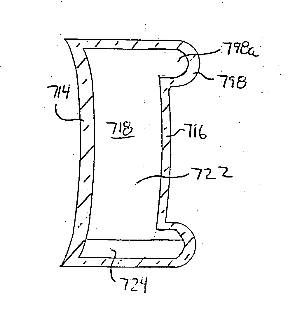
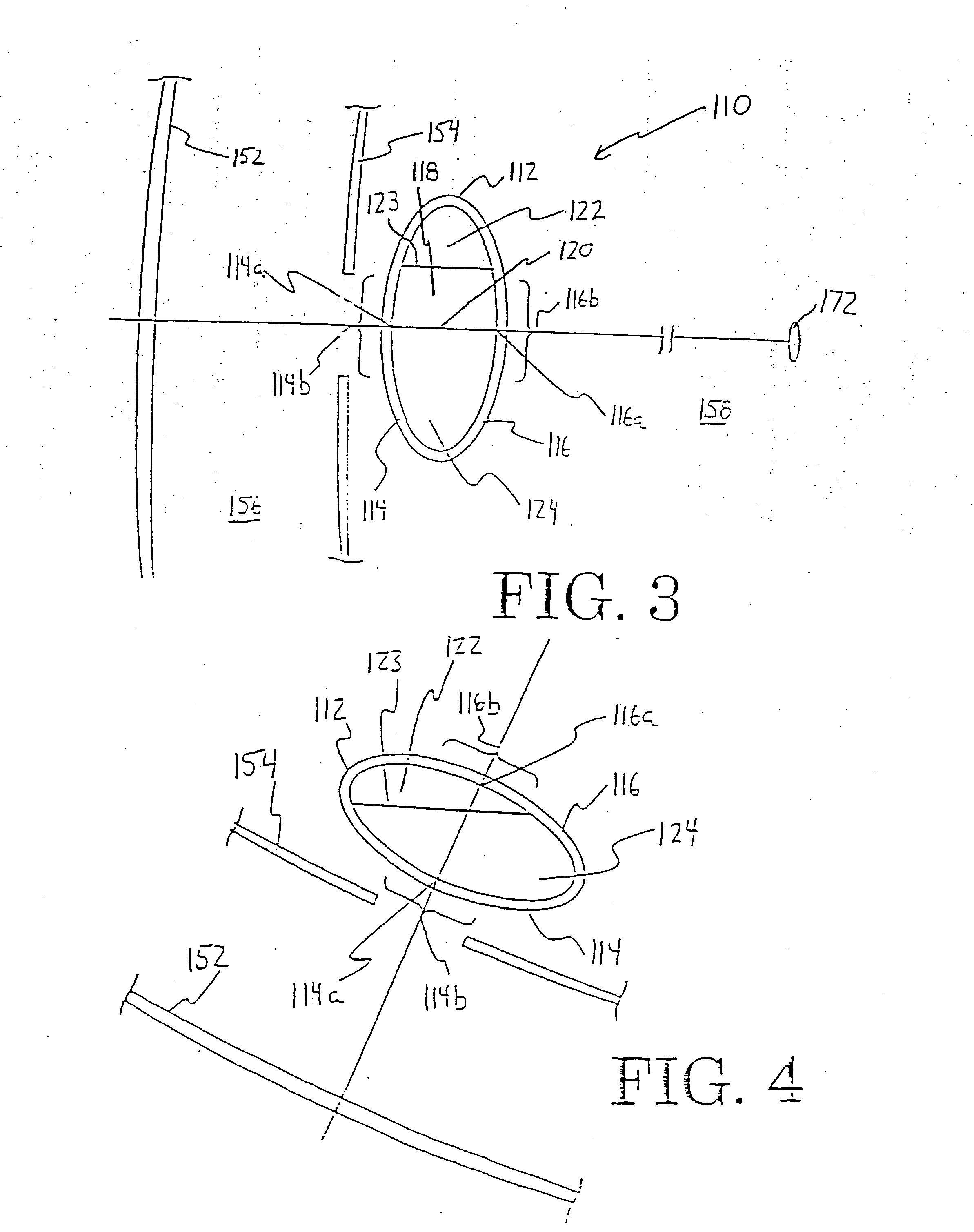
[0032] According to another embodiment of the first aspect of the invention, the primary fluid comprises an optically transmissive upper fluid, and is contained in the chamber of the optic body in a sufficient amount that orienting the optical axis in a horizontal orientation for far vision positions the optical axis through the upper fluid for immersing most of the anterior surface area of the anterior visual zone and most of the posterior surface area of the posterior visual zone in the upper fluid. Further, the secondary fluid comprises an optically transmissive lower liquid substantially immiscible with the upper fluid, and is contained in the chamber of the optic body below the upper fluid in a sufficient amount that orienting the optical axis for near vision at a range of effective downward angles relative to the horizontal orientation for near vision translates the lower liquid toward the anterior wall and positions the optical axis to extend through the lower liquid at the anterior optical center and the upper fluid at the posterior optical center for immersing most of the anterior surface area of the anterior visual zone with the lower liquid and most of the posterior surface area of the posterior visual zone in the upper fluid.
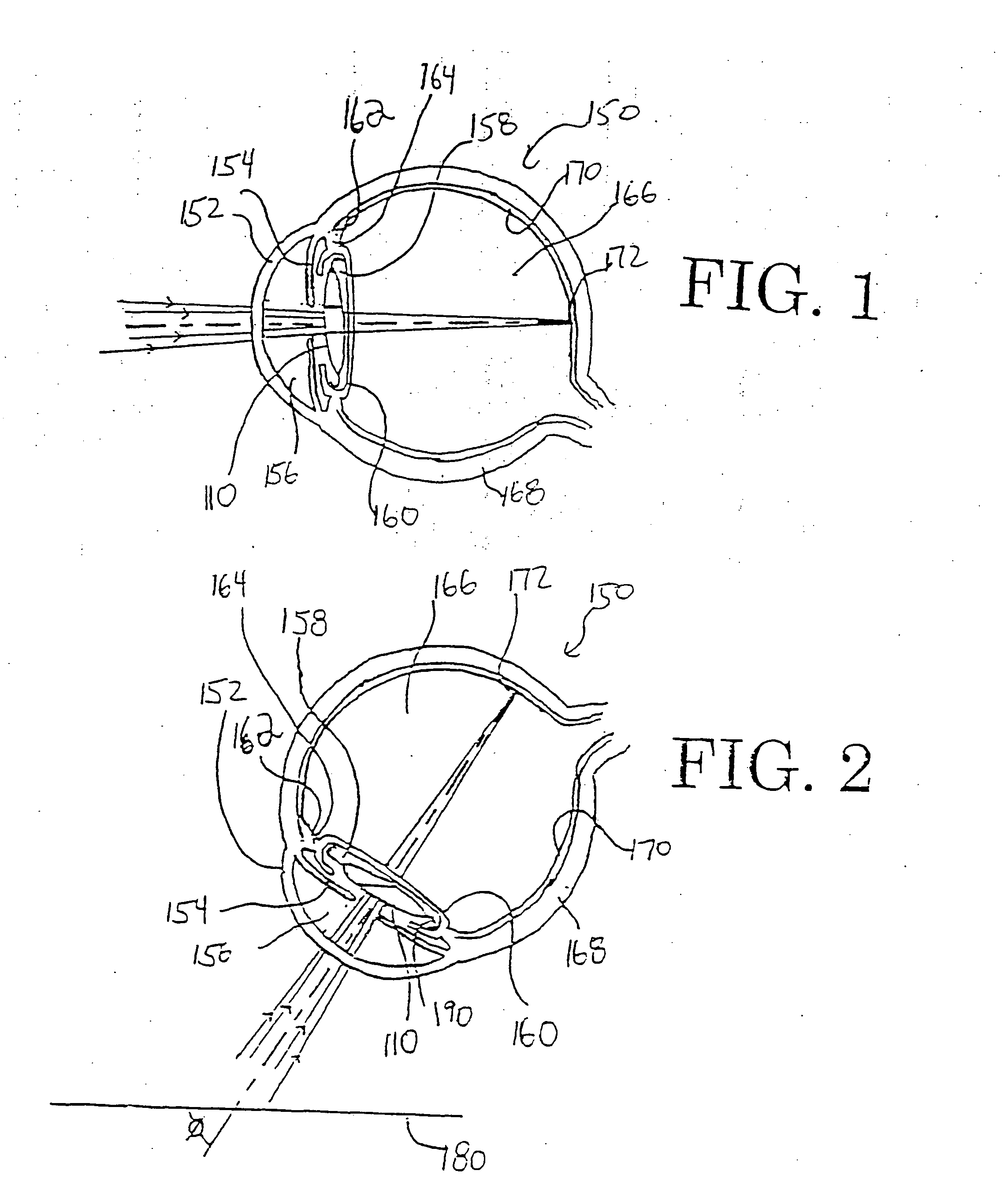
[0033] In accordance with the construction of the intraocular lens of this invention, multi-focus vision is achieved by the natural motion of the user's eye and / or head, preferably without requiring external visual correction devices, such as eyeglasses or contact lenses. For distant or far vision, the user gazes straight ahead to orient the optical axis substantially parallel to the horizon. In this straight-ahead gaze, the optical axis passes through either the optically transmissive lower liquid or the optically transmissive upper fluid. The refractive index of the fluid through which the optical axis passes and the curvature of the optic body alter the effective power of the lens for focusing for far distance (at the pr).
[0036] In another preferred embodiment of this invention, the adjustment in effective power of the lens is achieved without any moving parts (other than the flow of the refractive liquids) and without requiring the division of the intraocular lens into separate compartments via internal channels that prevent or inhibit elastic deformation of the lens.
[0038] In accordance with another aspect of this invention, a method is provided for using the intraocular lens of this invention. According to one preferred embodiment, an incision is created in the cornea, conjunctiva, and / or sclera of an eye having a posterior chamber and an anterior chamber. The intraocular lens is inserted into either the anterior chamber or posterior chamber of the eye through the incision. Preferably, the intraocular lens is placed in the posterior chamber of the eye, and more preferably the intraocular lens replaces a disposable lens in the capsular bag positioned posterior to the iris. The methods of this invention are especially useful for replacing a physiological lens that is partially or virtually totally defective, such as in the case of low vision and / or a cataractous lens. The methods of this invention also find utility in the replacement or supplementation of partially defective lenses, in addition to providing low vision for macular degeneration, aiding myopia and hyperopia and presbyopia, where glasses, contact lenses, or other corrective devices are needed for correcting the partial defect. The lens may also be used in a refractive correction and / or presbyopic surgical procedure.
[0039] A method according to another aspect of this invention comprises optically moving images away from a scotomatous area of a person having a retinal degenerative condition. The method comprises inserting an ocular lens into an eye of the person having the retinal degenerative condition, the ocular lens comprising an optic body, an optically transmissive primary fluid, and an optically transmissive secondary fluid. The optical body comprises an anterior wall, a posterior wall, and a chamber between the anterior wall and the posterior wall, the optically transmissive primary and secondary fluids contained in the chamber and having different densities and refractive indexes from one another. The ocular lens preferably is designed so that orienting the human eye in a generally straight ahead gaze for far vision passes the visual axis through the primary liquid, but not the secondary liquid, for focusing on a distant point, and moving the human eye into a downward gaze to pass the visual axis through the primary liquid and the secondary liquid for focusing on a near point. The method preferably further comprises providing an objective lens in front of the ocular lens. Preferably, the ocular lens is negative in power in straight ahead gaze and positive in power in down gaze, and the objective lens has a positive power. Collectively, the ocular and objective lenses provide a Galilean telescopic effect in straight ahead gaze and increased magnification in down gaze.

Problems solved by technology

According to another theory, the physiological lens enlarges with age causing a decrease in working distance between the ciliary body and the lens, resulting in decreased focus ability for the same muscle action.
The ciliary body-zonules-lens complex becomes less efficient at accommodating the focus of these rays on the retina.
Aphakia is the condition in which the crystalline lens is either absent or, in very rare cases, displaced from the pupillary area so that it adversely affects the eye's optical focusing system.
With advancing age, the physiological crystalline lens tends to develop opacities—a condition known as cataractogenesis—which unless treated eventually leads to blindness.
However, current synthetic intraocular lenses lack the flexibility of a physiological crystalline lens.
As a consequence, it is difficult, if not impossible, for the ciliary muscle to focus current synthetic intraocular lenses in the same way as a physiological lens to adjust for objects near the pp.
Thus, conventional monofocal intraocular lenses provide little, if any accommodating ability.
However, “reading” glasses and contact lenses have the drawbacks of being inconvenient, uncomfortable, susceptible to loss and breakage, and in the case of glasses, aesthetically undesirable to some users.
One problem with these designs is the zones of far and near are present simultaneously on the retina, thereby resulting in some blur or visual distortion at distance and near.
Attempts to create a “focusing” intraocular synthetic lens have been less than successful, and presbyopia, whether age-related or in pseudophakia, continues to be a vexing problem within eye care with no highly successful solutions yet in existence.
Another drawback to intraocular lenses is that an eye that has received an implant for restoring the natural accommodation of the eye may have its refractive error changed by the process of the implantation itself.
In this event, the focusable implant may function properly, but the powers needed to achieve clear distance focus may not be the same as calculated prior to insertion of the implant.
For example, an IOL selected for an eye of a particular length that is to be operated on might not function to fully correct maximum distance vision after the eye has healed.
As the eye heals, the lens may settle off-axis, tilt, or translate further forward or backward than the surgeon intended, leaving a refractive error that will make it necessary for the patient to use distance corrective lenses to see clearly.
First of all, the devices are heavy and bulky making them difficult to use from an ergonomic perspective.
Second, the devices, such as those mounted on a pair of spectacles, may be considered aesthetically unappealing by some.
Third, the devices may distort or decrease the effectiveness of magnification, for example, in the case of spectacle-mounted telescopic devices in which there exists a vertex distance (the distance from the back of the lens to the front of the cornea).
Fourth, the devices limit the user's mobility as the telescopic devices are usually housed in a casing that limits the user's peripheral vision.
Fifth, in the example of near vision magnification, the devices are often housed in a hand held device, which requires the user to not have “hands free” use of the device, i.e., the user may have trouble holding a newspaper in one hand and a device in the other.
Sixth, current implantable telescope lenses are held within bulky housings, which decrease the user's peripheral vision and result in a significant decrease in the user's mobility.

Method used


Image

  • Multi-focal intraocular lens, and methods for making and using same
  • Multi-focal intraocular lens, and methods for making and using same
  • Multi-focal intraocular lens, and methods for making and using same

Examples


examples

[0134] All examples were modeled on the Zemax Version 10.0 optical design program, SE edition, from Focus Software, Inc.

[0135] The human eye was first modeled as a typical or schematic adult human emmetrope, as described in the Optical Society of America Handbook. Each of the models described below is for a posterior chamber IOL design. The following assumptions were made for the human eye for the purposes of the calculations. The model was assumed to have spherical surfaces only (whereas the real cornea and lens are actually aspherics). Each structure of the schematic human eye was assumed to be made of a material having a uniform or homogenous index (whereas in the real human eye, the index of refraction may vary somewhat through each structure of the eye). The model also assumed that the capsular bag walls were very thin and parallel, i.e., non-existent. The lens was assumed to have symmetric radius, i.e., spherical. The pr was assumed to be 10 meters. Three wavelengths with equ...


PUM


Abstract

An intraocular lens is provided that includes an optic body having anterior and posterior walls, a chamber, and optically transmissive primary and secondary fluids, and method for making and using the same. One of the fluids is a gas. The secondary fluid is substantially immiscible with the primary fluid and has a different density and a different refractive index than the primary fluid. The primary fluid is present in a sufficient amount that orienting optical body optical axis horizontally for far vision positions the optical axis through the primary fluid, thereby immersing the anterior and posterior optical centers in the primary fluid. The secondary fluid is contained in the optic body in a sufficient amount that orienting the optical axis over a range of effective downward angles relative to the horizontal for near vision positions the optical axis to extend through the primary fluid and the secondary fluid, thus changing the focus of the intraocular lens.

Description

RELATED APPLICATIONS [0001] This application is a continuation of 10 / 733,173 filed Dec. 10, 2003, which is a continuation-in-part of PCT / US02 / 17964, filed Jun. 7, 2002, which is a continuation-in-part of U.S. patent application Ser. No. 10 / 158,574, filed in the U.S. Patent & Trademark Office on May 30, 2002, which is a continuation-in-part of U.S. patent application Ser. No. 10 / 139,144, filed in the U.S. Patent & Trademark Office on May 3, 2002, the complete disclosures of which are incorporated herein by reference. [0002] This application claims the benefit of priority of provisional patent application 60 / 297,306 filed in the U.S. Patent & Trademark Office on Jun. 11, 2001, the complete disclosure of which is incorporated herein by reference.BACKGROUND OF THE INVENTION [0003] 1. Field of the Invention [0004] This invention relates generally to bifocal and other multi-focal intraocular lenses, their production, and to their implantation and use in the eye. In particularly preferred ...

Claims


Application Information

Patent Timeline
Patent Type & Authority Applications(United States)
IPC IPC(8): A61F2/16A61F2/00A61F2/14A61F9/007
CPCA61F2/1613A61F2/1616A61F2/1618A61F2250/0053A61F2/1648A61F2250/0015A61F2/1624A61F2/1627
Inventor GLAZIER, ALAN N.WEBER, WILLIAM L.
Owner VISION SOLUTION TECH LLC
Why Eureka
Try Eureka