Afterglow dc-offset calibration in a nuclear imaging system

a nuclear imaging and afterglow technology, applied in the field of nuclear medicine, can solve the problems of inaccuracy of information, and inability to accurately calculate the integrated value of the even

Active Publication Date: 2007-03-29
SIEMENS MEDICAL SOLUTIONS USA INC
View PDF1 Cites 2 Cited by
  • Summary
  • Abstract
  • Description
  • Claims
  • Application Information

AI Technical Summary

Benefits of technology

[0016] The present invention solves the existing need according to a first aspect by providing a method of calibrating a nuclear medical imaging apparatus for effects of scintillation pulse afterglow, including the steps of determining a minimum value of an Esum output signal of the apparatus in response to a relatively low count rate radiation source; determining a minimum value of an Esum output signal of the apparatus in response to a relatively high count rate radiation source; c) comparing the minimum value of the high count rate Esum output signal with the minimum va

Problems solved by technology

Another instance of inaccurate information may arise when two gamma photons interact with the scintillation crystal within a time interval that is shorter than the time resolution of the system (in other words the amount of time required for a light event to decay sufficiently such that the system can process a subsequent light event as an independent event), such that light events from the two gamma interactions are said to “pile up,” or be superposed on each other.
The signal resulting from a pulse pile-up would be meaningless, as it would not be possible to know whether the pulse resulted from two valid events, two invalid events, or one valid event and one invalid event.
Where there are many gamma interactions in a period of time that is short as compared with the longer decay time constant, the accumulated value of the slowly decaying tails of many prior events gets added to the integrated value of the current event being process

Method used


Image

  • Afterglow dc-offset calibration in a nuclear imaging system
  • Afterglow dc-offset calibration in a nuclear imaging system
  • Afterglow dc-offset calibration in a nuclear imaging system

Examples


Embodiment Construction

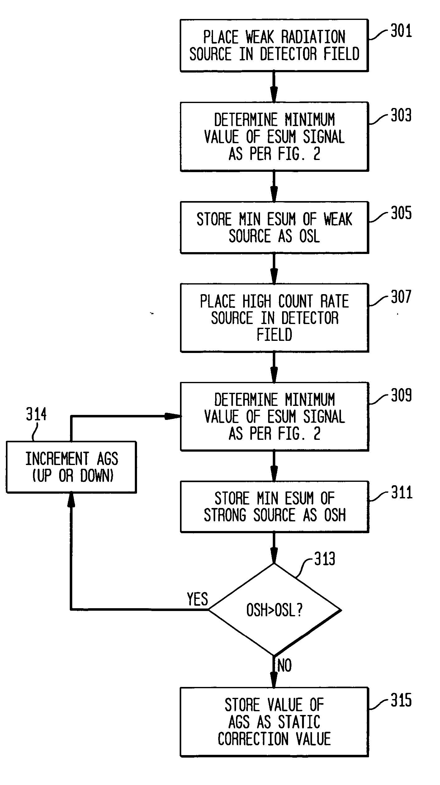
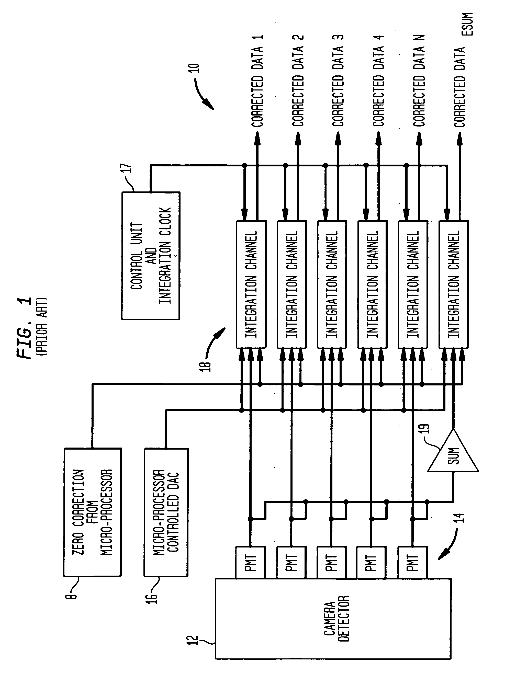
[0024] Referring to FIG. 1, a medical imaging system 10 according to the prior art, as disclosed in the aforementioned '395 patent, produces signals corresponding to events detected by a sensor such as a scintillation crystal camera detector 12. Upon absorption of incident gamma rays in the detector 12, corresponding scintillation light events are amplified by a plurality of photodetectors such as PMTs (photomultiplier tubes) 14 forming a two-dimensional array on the scintillation crystal of the detector 12. Each PMT corresponds to a respective channel, the signals from which are processed together with signals from the other channels, to calculate the spatial location of a gamma event occurring in the scintillation crystal.

[0025] The signals from the plurality of PMT channels are processed by a plurality of integration channel processing units 18 incorporating signal processing circuitry, with each processing unit 18 being operatively connected to a corresponding one of PMTs 14 fo...


PUM


Abstract

Method and system are provided for calibrating a nuclear medical imaging apparatus for DC shift caused by gamma event afterglow pulses. Detector responses to weak and to high count rate radiation sources are compared with each other, and a zero correction value is incremented until the detector response is the same for the weak source and the high count rate source. The zero correction value is then stored as a static zero correction value, which multiplies a dynamic zero correction value obtained just prior to the occurrence of a gamma event, in order to remove the effects of DC shifts from the output energy signal Esum.

Description

BACKGROUND OF THE INVENTION [0001] 1. Field of the Invention [0002] The present invention generally relates to nuclear medicine, and systems for obtaining nuclear medicine images of a patient's body organs of interest. In particular, the present invention relates to a novel method and system for more accurately detecting the occurrence of valid scintillation events. [0003] 2. Description of the Background Art [0004] Nuclear medicine is a unique medical specialty wherein radiation is used to acquire images that show the function and anatomy of organs, bones or tissues of the body. Radiopharmaceuticals are introduced into the body, either by injection or ingestion, and are attracted to specific organs, bones or tissues of interest. Such radiopharmaceuticals produce gamma photon emissions that emanate from the body. One or more detectors are used to detect the emitted gamma photons, and the information collected from the detector(s) is processed to calculate the position of origin of t...

Claims


Application Information

Patent Timeline
IPC IPC(8): G01T1/161
CPCG01T1/1642G01T1/171A61B6/4258G01T7/005G01T1/208
Inventor ARSENEAU, ROGER E.
Owner SIEMENS MEDICAL SOLUTIONS USA INC
Why Eureka
Try Eureka