Nuclear medicine imaging apparatus and a method for generating image data

Active Publication Date: 2007-05-03
TOSHIBA MEDICAL SYST CORP
View PDF4 Cites 19 Cited by
  • Summary
  • Abstract
  • Description
  • Claims
  • Application Information

AI Technical Summary

Benefits of technology

[0035] According to a nuclear medicine imaging apparatus and a method for generating image data consistent with the present invention, when projection data are generated based on count values emitted from an object (patient) who has been administered with a radioactive isotope, it become possible to body moveme

Problems solved by technology

For instance, if it is required s to move the detector by 10 steps for collecting projection data for a required diagnosis scope, it takes more than twenty minutes.
Consequently, the patient may move his body during such a long data collecting time for a wide diagnosis area of an object.
When a patient moves his body during such a data collecting time (hereinafter, simply, “a body movement “has occurred), a problem in the image quality of image data being generated based on such projection data conventionally occurred.
Thus, image quality deteriorates due to

Method used


Image

  • Nuclear medicine imaging apparatus and a method for generating image data
  • Nuclear medicine imaging apparatus and a method for generating image data
  • Nuclear medicine imaging apparatus and a method for generating image data

Examples


Embodiment Construction

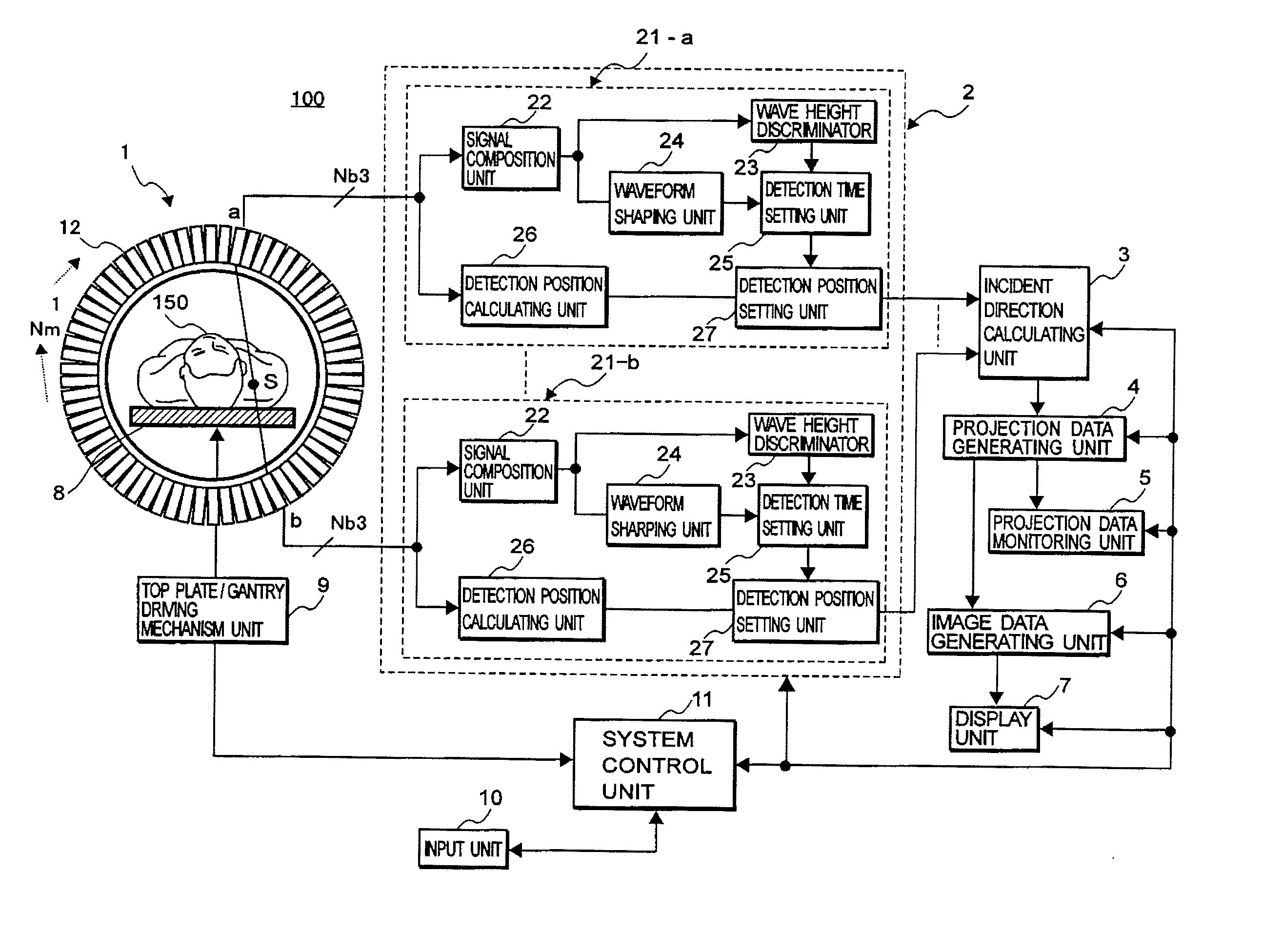
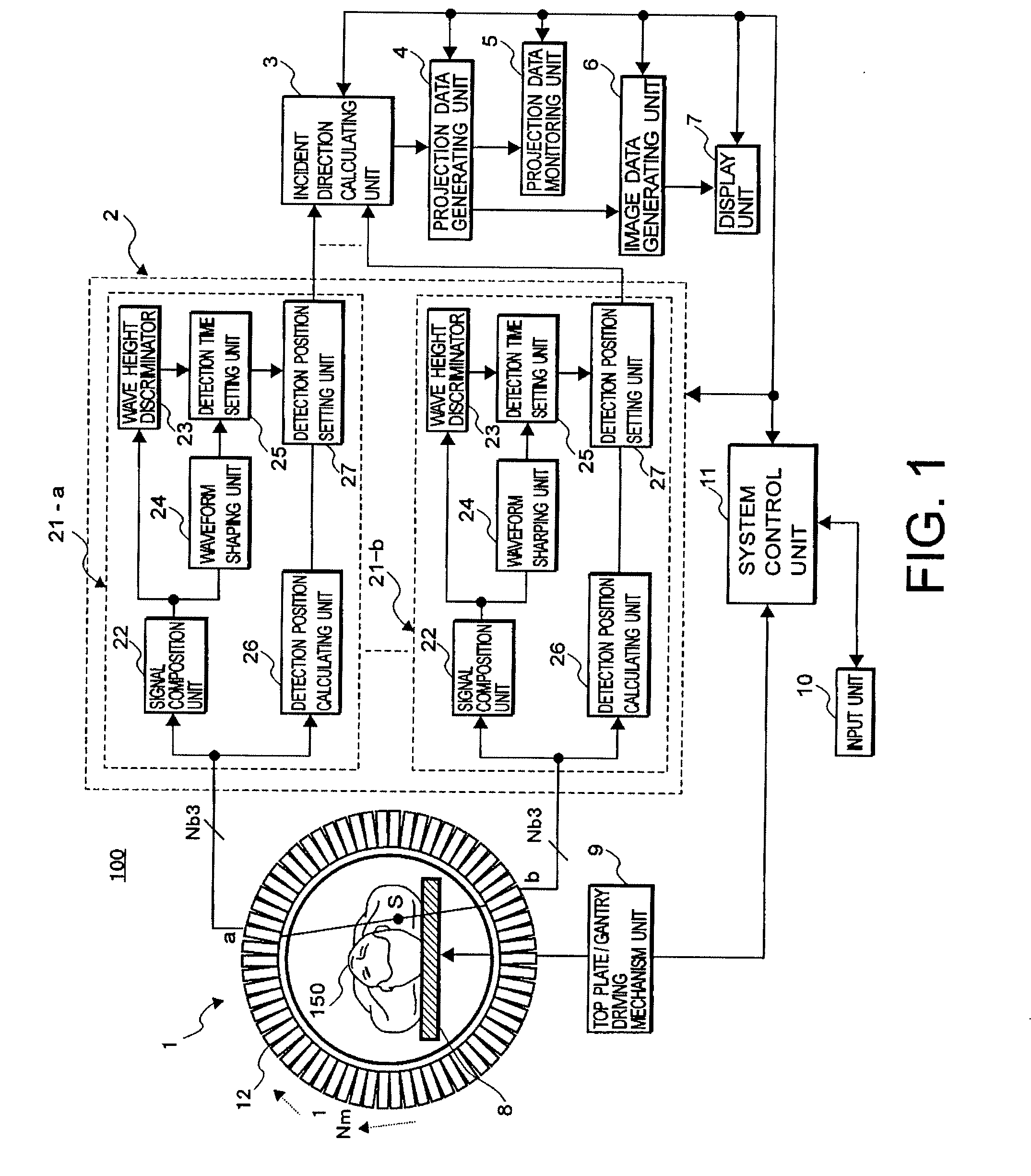
[0045] With reference to the following FIGS. 1-8, embodiments consistent with the present invention will be explained. As an exemplary embodiment according to the nuclear medicine imaging apparatus consistent with the present invention, it will be explained as to a PET apparatus. Of course, the present invention is applicable to nuclear medicine imaging apparatus for other image diagnosis apparatus, such as a SPECT apparatus or a composite type PET apparatus with an X-ray CT apparatus.

[0046] An object (patient) has previously been administered (injected) a radioactive isotope before being positioned into a nuclear medicine imaging apparatus consistent with the present invention. In the nuclear medicine imaging apparatus, during a prescribed monitoring time period, a pair of gamma-rays emitted from a diagnostic portion in the patient with the radioactive isotope is successively detected by a detector of a ring-like configuration that is comprised of a plurality of detector modules. ...


PUM


Abstract

The present invention provides a nuclear medicine imaging apparatus and image data generation method that achieves restarting of the generation of projection data and at an early stage while monitoring a variation of count values for detecting an occurrence of non-permissible body movement of a patient. The image processing apparatus consistent with the present invention detects a pair of gamma-rays successively emitted from an object with a radioactive isotope through a pair of detector modules in a data detecting unit. A data processing unit and an incident direction calculating unit in the image processing apparatus respectively calculate a gamma-ray detection position and a gamma-ray incident direction based on the acquired detection signals. A projection data generating unit in the apparatus generates monitoring projection data based on each count value of the detection signals in correspondence to the gamma-ray detection position and the gamma-ray incident direction. A projection data monitoring unit calculates a body movement index of the object by comparing count values of the monitoring projection data that are generated in each of two preferably adjoining monitoring periods. A system control unit generates an alarm signal for performing repetition of the monitoring projection data when the body movement index exceeds a threshold value and displays the alarm signal on a display unit.

Description

CROSS-REFERENCE TO RELATED APPLICATIONS [0001] This application claims priority from, and the benefit of, Japanese Patent Application No. 2005-298717, filed on Oct. 13, 2004, the contents of which are expressly incorporated herein by reference in their entirety. BACKGROUND OF THE INVENTION [0002] A. Field of the Invention [0003] The present invention relates to a nuclear medicine imaging apparatus and a method for displaying image data thereof, and more particularly, to a positron emission computer tomography (PET) apparatus and a method for displaying image data thereof that can automatically restart data collection of image data by detecting impermissible body movement of an object during the image data collection. [0004] B. Background of the Invention [0005] Recent developments of computer technologies have rapidly progressed medical imaging apparatus, such as an X-ray diagnosis apparatus, a magnetic resonance imaging (MRI) apparatus, an X-ray computer tomography (CT) apparatus a...

Claims


Application Information

Patent Timeline
IPC IPC(8): G01T1/164G01T1/161
CPCG01T1/1642G01T1/1648G01T1/2985
Inventor TANAKA, TAKASHI
Owner TOSHIBA MEDICAL SYST CORP
Why Eureka
Try Eureka