Antibodies, assays and kits to quantitate cartilage destruction

Inactive Publication Date: 2007-05-03
SANDY JOHN D +2
View PDF58 Cites 4 Cited by
  • Summary
  • Abstract
  • Description
  • Claims
  • Application Information

AI Technical Summary

Benefits of technology

[0022] In another embodiment, the method further involves, prior to contacting, administering to the subject an agent for treating a cartilage destruction condition, such that the amount of epitope is a measure of prevention of progressive cartilage destruction.

Problems solved by technology

Many current methods for determining presence and rate of cartilage destruction during arthritic processes are invasive, or are associated with highly technological and expensive equipment and are performed only at specialized centers.

Method used


Image

  • Antibodies, assays and kits to quantitate cartilage destruction

Examples


example 1

Preparation of Antibody Specific for a Cartilage Aggrecan Cleavage Site

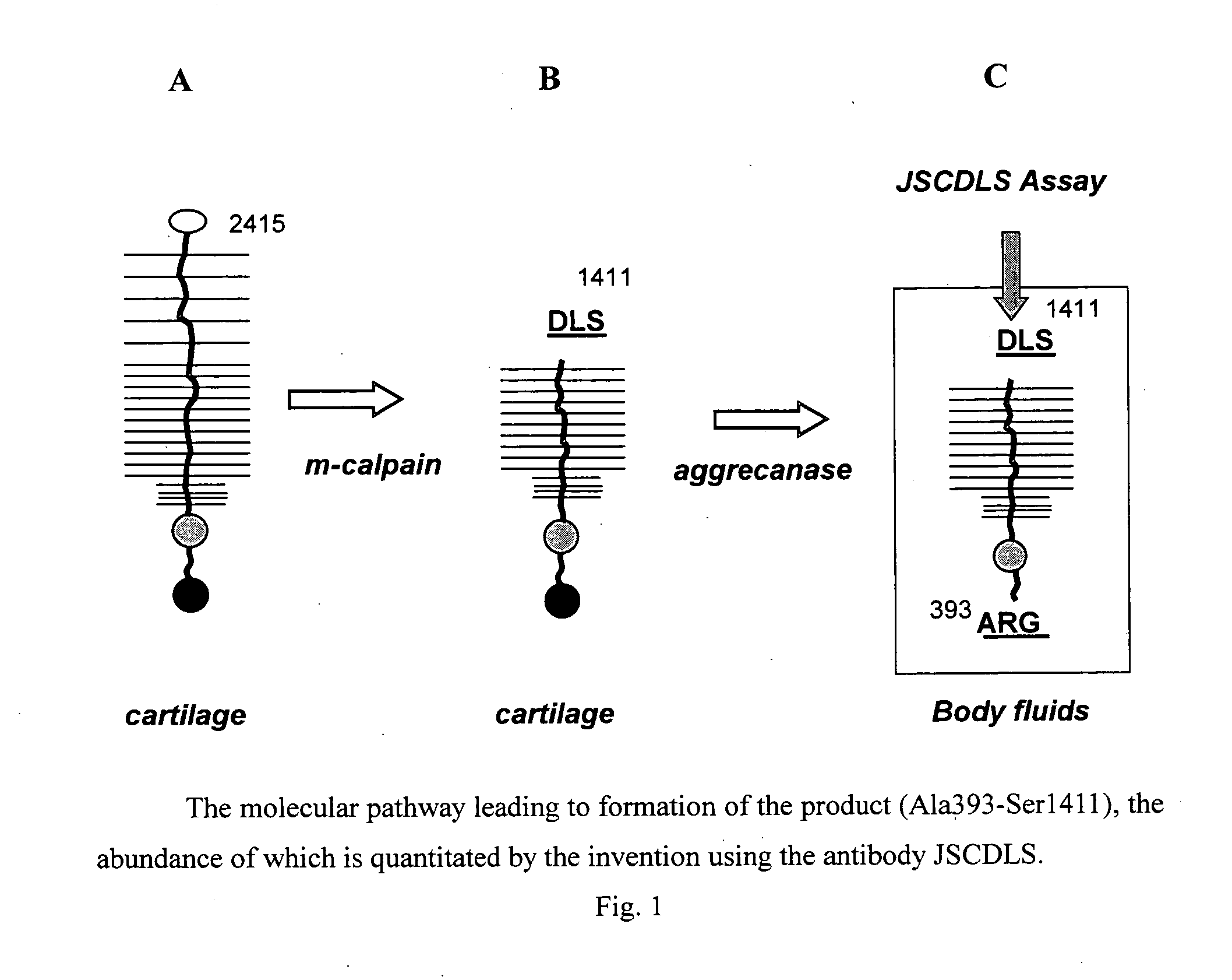
[0190] Aggrecanase attacks the stable intercellular matrix and generates both Ala393-Glu1564 and the “target” Ala393-Ser1411. The specific target is generated by aggrecanase-mediated cleavage of a pre-existing calpain product (see FIG. 1). The sequences of the junctions of the target have been determined herein, and these sequences used to develop an antibody that is specific for the epitope created by digestion by aggrecanase.

[0191] Antibody was produced by immunizing an animal, by contacting it with a synthetic peptide having amino acid sequence cys-gly-gly-ser-gly-val-glu-asp-leu-ser (SEQ ID NO: 1). Data showed that the antibody obtained recognizes the target peptide, i.e., is a ligand of this target.

[0192] The antibody was obtained from immunized rabbits, is identified herein as JSCDLS, and was used to coat wells of a multi-well dish, at various dilutions. To serve a positive control for antibody binding, ...

example 2

Correlating Target with Arthritic Conditions

[0193] Samples of body fluids are obtained from human subjects, in two groups: patients suffering with arthritic conditions, and normal subjects, are screened along with the dilutions of the target. In broad outline, volumes of each sample dilution are added to separate wells of the multi-well plate previously coated with JSCDLS antibody. Target molecules carrying the DLS epitope are immobilized to the antibody coated to the wells, and the wells are evacuated so that remaining volume of sample with unbound material is discarded, and the wells are washed. The target bound to the antibody is detected calorimetrically by one of numerous methods known to those of skill in the art of immunology (see Harlow, E. et al., 1988, Antibodies: a Laboratory Manual, Cold Spring Harbor Press, Cold Spring Harbor, N.Y.).

[0194] Samples from arthritic patients are found to contain significantly larger amounts of target, as determined by binding to the immob...

example 3

Pretreatment of Patient Samples Prior to Antibody Binding

[0195] In practice, it is convenient to pre-treat the samples, e.g., to trypsinize, and to digest with chondroitinase and neuraminidase, the target-containing fluids (to make disaccharide-substituted Ileu925-Ser1411). Samples are then reacted in wells of a multi-well dish, each well having been coated with antibody JSCDLS, to bind resulting further digested target Ileu925-Ser1411 to the antibody and then detect the “sandwich” peptide with peanut agglutinin peroxidase.

[0196] By any of the above techniques, target is assayed in various body fluids, most conveniently and least invasively being urine, and in synovial fluids.


PUM

PropertyMeasurementUnit
Solubility (mass)aaaaaaaaaa
Chemiluminescenceaaaaaaaaaa
Degradation propertiesaaaaaaaaaa

Abstract

Antibody compositions specific for peptides released during arthritic cartilage destruction are provided as reagents for assay of progression of arthritic processes for antibodies, and as diagnostic and prognostic agents. Also provided are methods and kits for this assay.

Description

RELATED APPLICATIONS [0001] The present application claims the benefit of provisional application Ser. No. 60 / 732,976 filed in the U.S. Patent and Trademark Office on Nov. 3, 2005, which is hereby incorporated by reference herein in its entirety.TECHNICAL FIELD [0002] The present invention relates to antibody compositions, assays and kits to quantitate cartilage destruction. BACKGROUND [0003] Arthritic diseases are ubiquitous in an aging population, with costs in 1991 of knee replacements alone due to osteoarthritis exceeding one billion dollars. Many current methods for determining presence and rate of cartilage destruction during arthritic processes are invasive, or are associated with highly technological and expensive equipment and are performed only at specialized centers. These methods include digital imaging analysis of microscopic images of biopsied tissue in experimental animals, and for human patients, include CAT scans (CT), magnetic resonance imaging (MR), and ultrasound...

Claims


Application Information

Patent Timeline
IPC IPC(8): G01N33/567C12P21/04C07K16/40
CPCC07K16/18C07K2317/34G01N33/574G01N2500/00G01N2800/105
Inventor SANDY, JOHN D.MASUDA, KOICHIPFISTER, BRIAN
Owner SANDY JOHN D
Why Eureka
Try Eureka