
Clip-Based Systems and Methods for Treating Septal Defects

a technology of septal defect and clip, applied in the field of clips for treating internal tissue defects, can solve the problems of high risk, serious complications for patients, and inherently difficult treatment of internal tissue defects

Inactive Publication Date: 2007-06-07
OVALIS
View PDF99 Cites 28 Cited by
  • Summary
  • Abstract
  • Description
  • Claims
  • Application Information

AI Technical Summary

Benefits of technology

"This patent describes improved devices, systems, and methods for closing internal tissue defects, such as septal defects. The invention involves delivering a clip with a tubular body into a hole in the septal wall, where the clip has two deflectable members that are positioned on either side of the wall. This results in the clip closing off the defect tunnel in the wall, maintaining it in a partially closed state. The technical effect of this invention is to provide a minimally invasive and effective treatment for heart defects."

Problems solved by technology

By nature of their location, the treatment of internal tissue defects is inherently difficult.
Access to a defect through invasive surgery introduces a high level of risk that can result in serious complications for the patient.
Access to the defect remotely with a catheter or equivalent device is less risky, but treatment of the defect itself is made more difficult given the limited physical abilities of the catheter.
The difficulty in accessing and treating tissue defects is compounded when the defect is found in or near a vital organ.
Occasionally, however, these foramen fail to close and create hemodynamic problems, which, in extreme cases, can prove fatal.
A PFO can pose serious health risks for the individual, including strokes and migraines.
These devices suffer from numerous shortcomings.
For instance, these devices typically involve frame structures that often support membranes, either of which may fail during the life of the patient, thereby introducing the risk that the defect may reopen or that portions of the device could be released within the patient's heart.
These devices can fail to form a perfect seal of the septal defect, allowing blood to continue to shunt through the defect.
Also, the size and expansive nature of these devices makes safe withdrawal from the patient difficult in instances where withdrawal becomes necessary.
The presence of these devices within the heart typically requires the patient to use anti-coagulant drugs for prolonged periods of time, thereby introducing additional health risks to the patient.
Furthermore, these devices can come into contact with other portions of the heart tissue and cause undesirable side effects such as an arrhythmia, local tissue damage, and perforation.

Method used


Image

  • Clip-Based Systems and Methods for Treating Septal Defects
  • Clip-Based Systems and Methods for Treating Septal Defects
  • Clip-Based Systems and Methods for Treating Septal Defects

Examples


Embodiment Construction

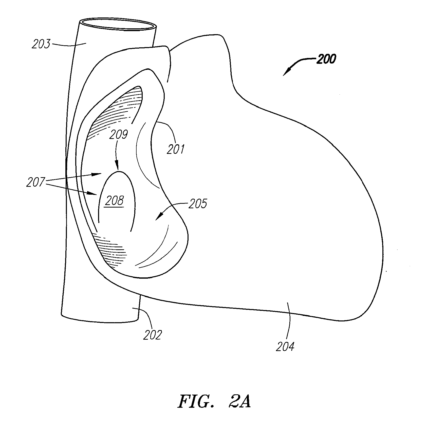
[0098] Deformable clip-type devices for treating internal tissue defects are described herein, along with systems for delivery of those devices as well as methods for using the same. For ease of discussion, these devices, systems and methods will be described with reference to treatment of a PFO. However, it should be understood that these devices, systems and methods can be used in treatment of any type of septal defect including ASD's, VSD's and the like, as well as PDA's, pulmonary shunts or other structural cardiac or vascular defects or non-vascular defects, and also any other tissue defect including non-septal tissue defects.

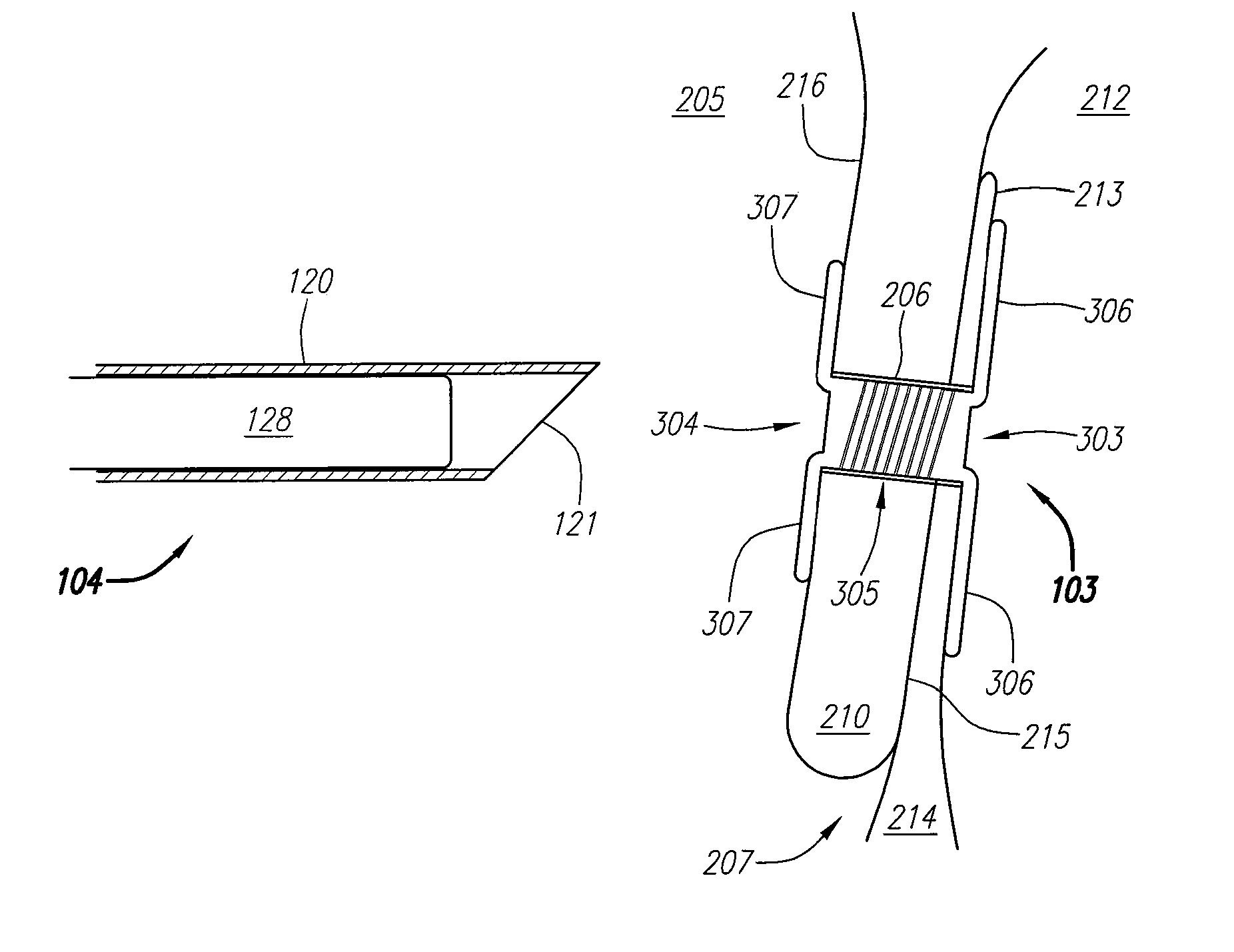
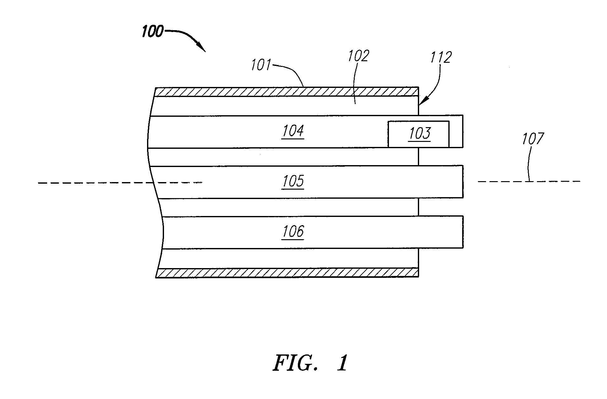
[0099]FIG. 1 is a block diagram depicting a distal portion of an exemplary embodiment of a septal defect treatment system 100 configured to treat and preferably close a PFO. In this embodiment, treatment system 100 includes an elongate body member 101 configured for insertion into the vasculature of a patient (human or animal) having a septal defect. Body...


PUM


Abstract

Systems and methods for treating internal tissue defects, such as septal defects, with clip-based devices are provided. An exemplary clip-based device includes a tubular body having at least a first and a second deflectable member coupled thereto. The first and second members are coupled on opposite ends of the tubular body and configured to deflect between an undeployed configuration and a deployed configuration. In the deployed configuration, each member extends outwardly away from the tubular body in a position configured to abut a tissue surface. The first and second members are preferably configured to maintain a tissue wall therebetween and at least partially close any opening in the tissue wall.

Description

CROSS-REFERENCE TO RELATED APPLICATIONS [0001] This application is a divisional of U.S. patent application Ser. No. 11 / 295,338, filed on Dec. 5, 2005, which is fully incorporated herein by reference.FIELD OF THE INVENTION [0002] The present invention relates generally to clips for treating internal tissue defects, such as septal defects, and systems and methods for delivering the same. BACKGROUND OF THE INVENTION [0003] By nature of their location, the treatment of internal tissue defects is inherently difficult. Access to a defect through invasive surgery introduces a high level of risk that can result in serious complications for the patient. Access to the defect remotely with a catheter or equivalent device is less risky, but treatment of the defect itself is made more difficult given the limited physical abilities of the catheter. The difficulty in accessing and treating tissue defects is compounded when the defect is found in or near a vital organ. For instance, a patent forame...

Claims


Application Information

Patent Timeline
Patent Type & Authority Applications(United States)
IPC IPC(8): A61B17/08
CPCA61B17/0057A61B2018/00392A61B17/221A61B2017/00247A61B2017/00575A61B2017/00579A61B2017/00592A61B2017/00606A61B2017/00615A61B2017/00619A61B2017/00623A61B2017/00867A61B2017/00893A61B2017/0412A61B2017/0419A61B2017/0422A61B2017/22047A61B2017/2926A61B2017/306A61B17/12122
Inventor ABBOTT, RYANBELEF, W. MARTINCARSON, DEANFITZGERALD, PETERJABBA, RONALD J.NIELSEN, JAMESTHORNTON, PETER
Owner OVALIS
Learn More