Method and apparatus for visually supporting an electrophysiological catheter application in the heart by means of bidirectional information transfer

a catheter application and electrophysiological technology, applied in the field of electrophysiological catheter application visual support, to achieve the effect of rapid overview of position and exten

Inactive Publication Date: 2007-07-19
SIEMENS AG
View PDF10 Cites 36 Cited by
  • Summary
  • Abstract
  • Description
  • Claims
  • Application Information

AI Technical Summary

Benefits of technology

[0007] The object of the present invention is to specify a method and an apparatus for visually supporting an electrophysiological catheter

Problems solved by technology

The suboptimal operator orientation possibilities for guiding the catheter cons

Method used


Image

  • Method and apparatus for visually supporting an electrophysiological catheter application in the heart by means of bidirectional information transfer
  • Method and apparatus for visually supporting an electrophysiological catheter application in the heart by means of bidirectional information transfer
  • Method and apparatus for visually supporting an electrophysiological catheter application in the heart by means of bidirectional information transfer

Examples


Embodiment Construction

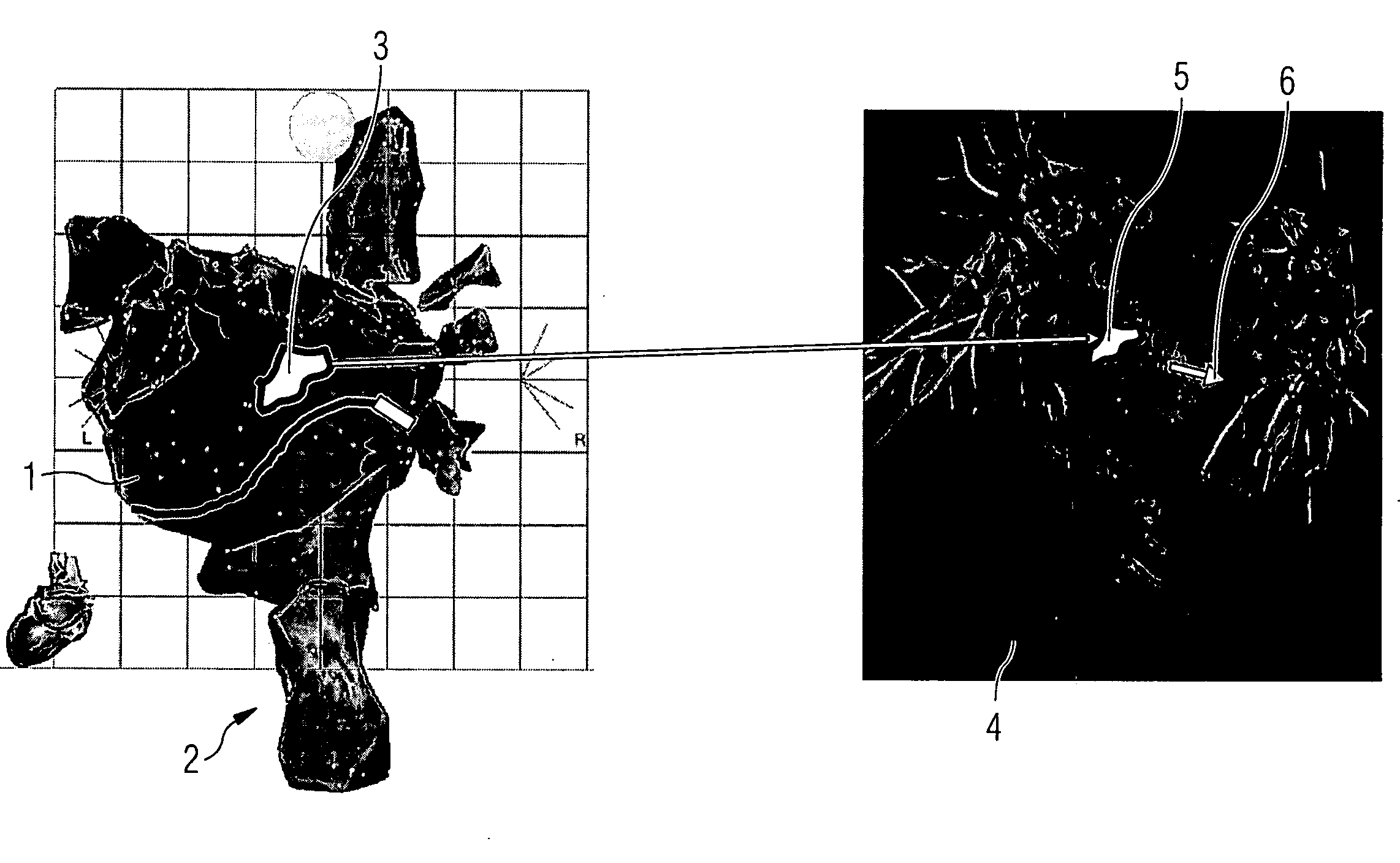
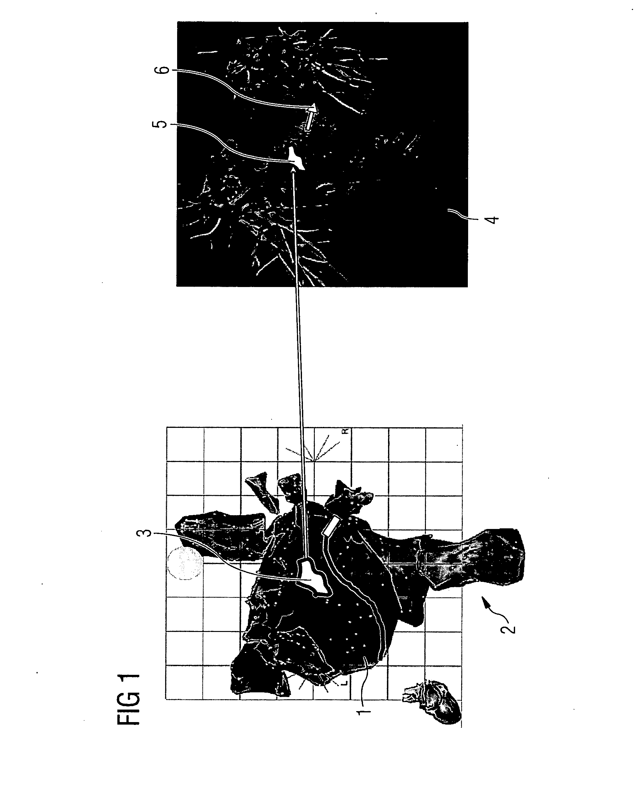
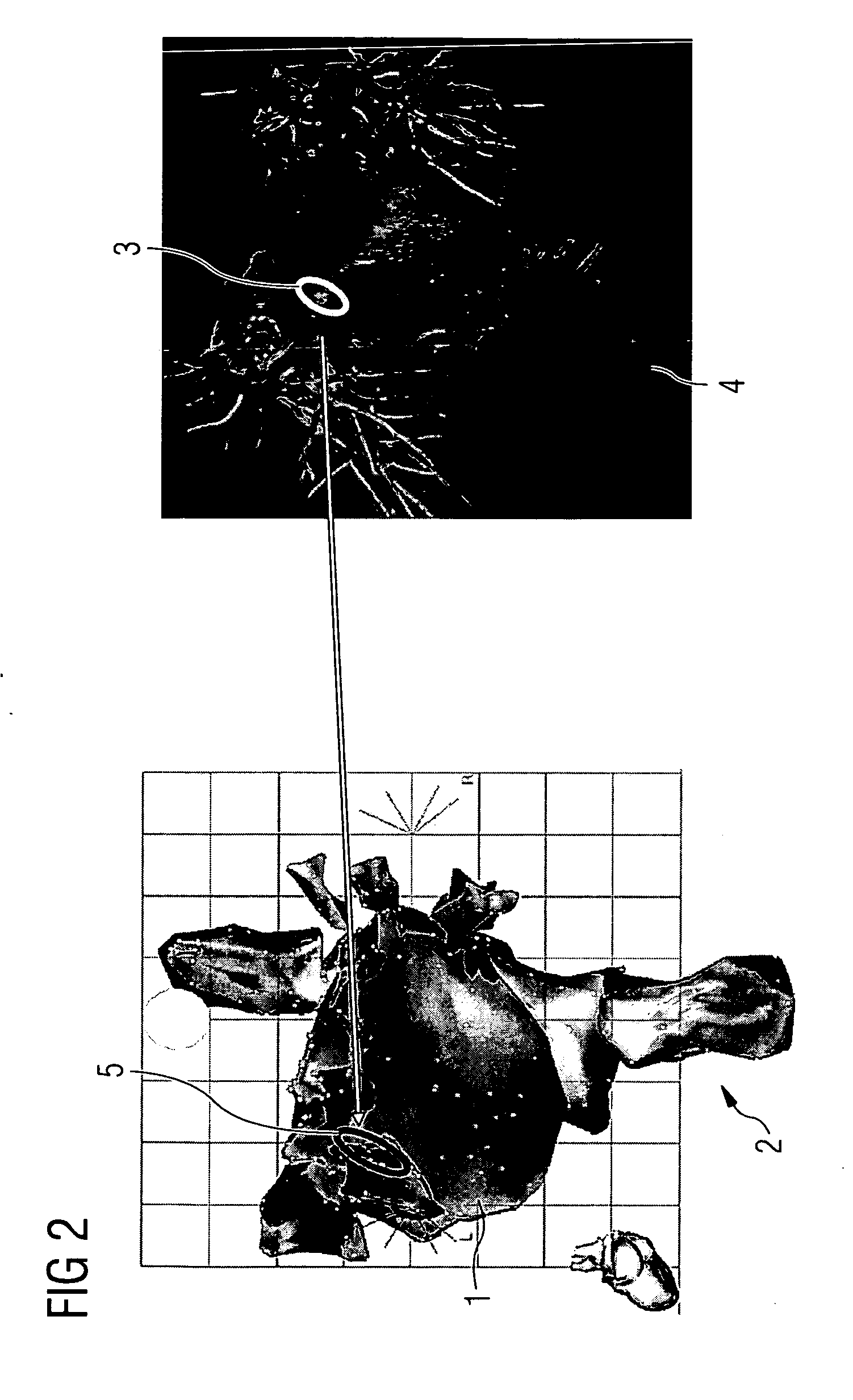
[0023]FIG. 1 shows an example of transferring the contour of an area 3 captured in the electroanatomical mapping data from the electroanatomical mapping system to the 3D visualization workstation on which a representation 4 of the 3D image data is displayed. During the electrophysiological procedure, an electroanatomical 3D map 2, as shown in the left-hand part of FIG. 1, is visualized on the electroanatomical 3D mapping system. This representation is based on a simplified model of the heart on which the individual 3D mapping data are visible as mapping points 1. In this representation the position and orientation of the mapping catheter 9, which can be derived from the 3D mapping data, is additionally superimposed. In this 3D mapping data, the position and contour of a three-dimensional area 3 is now captured which is visible in the left-hand part of the figure, this possibly being, for example, the outline of post-infarction scarring of the left ventricle which can be determined f...


PUM


Abstract

Method and apparatus for visually supporting an electrophysiological catheter application in the heart by means of bidirectional information transfer
The present invention relates to a method and apparatus for visually supporting an electrophysiological catheter application in the heart. For the method, 3D image data of at least the heart, which is captured using a tomographic 3D imaging method prior to execution of the catheter application, and electroanatomical 3D mapping data of at least one area of the heart to be treated, which is captured during execution of the catheter application, is provided and the electroanatomical 3D mapping data and/or at least part of the 3D image data is displayed during execution of the catheter application. The method is characterized in that, in the electroanatomical 3D mapping data and/or the 3D image data, the contour of one or more areas (3) relevant to the catheter application is captured and transferred to the other system in each case on which the areas (3) are superimposed as a single polyline (5) in the representation (2, 4) of the electroanatomical 3D mapping data and/or 3D image data. The method proposed and the associated apparatus provide the user with a rapid overview of the areas relevant to the catheter application.

Description

CROSS REFERENCE TO RELATED APPLICATIONS [0001] This application claims priority of German application No. 10 2005 042 329.9 filed Sep. 06, 2005, which is incorporated by reference herein in its entirety. FIELD OF THE INVENTION [0002] The present invention relates to a method and apparatus for visually supporting an electrophysiological catheter application in the heart, in which 3D image data of at least the heart, which is captured using a tomographic 3D imaging method prior to execution of the catheter application, and electroanatomical 3D mapping data of at least one area of the heart to be treated, which is captured during execution of the catheter application, is provided and the electroanatomical 3D mapping data and / or at least part of the 3D image data is displayed during execution of the catheter application. BACKGROUND OF THE INVENTION [0003] The treatment of cardiac dysrhythmias has changed significantly since the introduction of the technique of catheter ablation by means...

Claims


Application Information

Patent Timeline
IPC IPC(8): A61B5/05
CPCA61B18/1492A61B19/52G06T11/008A61B2018/00839A61B2019/5289A61B2017/00053A61B90/36A61B2090/364
Inventor BOESE, JANMEYERPFISTER, MARCUSRAHN, NORBERT
Owner SIEMENS AG
Why Eureka
Try Eureka