Digital medical image processing

a medical image and digital technology, applied in image data processing, character and pattern recognition, instruments, etc., can solve the problems of time-consuming and laborious processing and perform the task with great car

Inactive Publication Date: 2007-09-13
MEDICSIGHT
View PDF3 Cites 6 Cited by
  • Summary
  • Abstract
  • Description
  • Claims
  • Application Information

AI Technical Summary

Benefits of technology

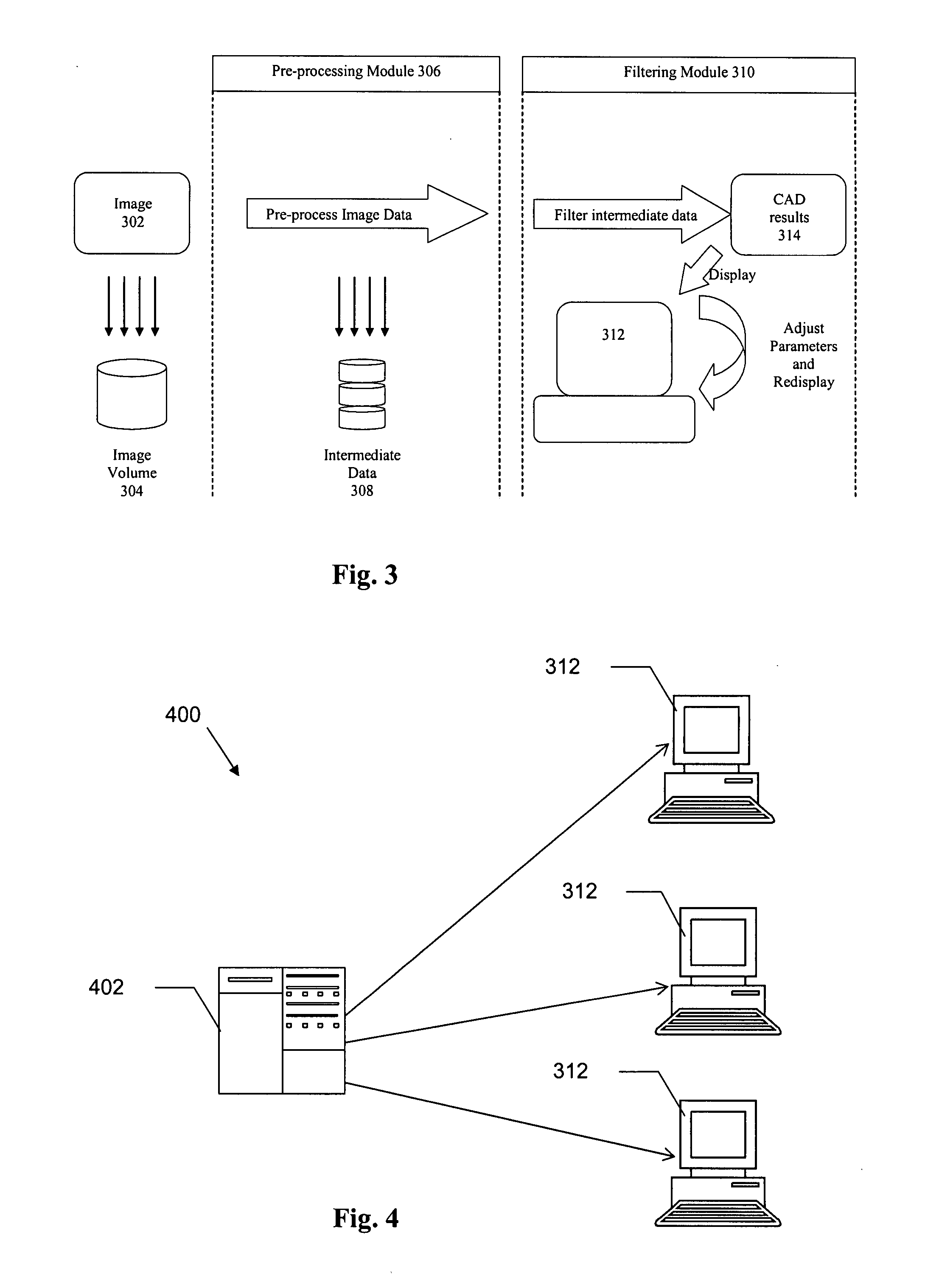
[0014]An advantage of embodiments of the invention is that they allow distributed CAD analysis. For example, processing-intensive earlier stages may be performed by medical image servers, and the results communicated to viewer workstations so that the final CAD results may be customized without ...

Problems solved by technology

However, the process is time consuming and must be per...

Method used


Image

  • Digital medical image processing
  • Digital medical image processing
  • Digital medical image processing

Examples


Embodiment Construction

[0024]This specification discloses one or more embodiments that incorporate the features of this invention. The embodiment(s) described, and references in the specification to “one embodiment”, “an embodiment”, “an example embodiment”, etc., indicate that the embodiment(s) described may include a particular feature, structure, or characteristic, but every embodiment may not necessarily include the particular feature, structure, or characteristic. Moreover, such phrases are not necessarily referring to the same embodiment. Furthermore, when a particular feature, structure, or characteristic is described in connection with an embodiment, it is submitted that it is within the knowledge of one skilled in the art to effect such feature, structure, or characteristic in connection with other embodiments whether or not explicitly described.

I. MEDICAL IMAGING

[0025]The present invention is applicable to digital medical images. One example of such an image is a computed tomography (CT) scan im...


PUM


Abstract

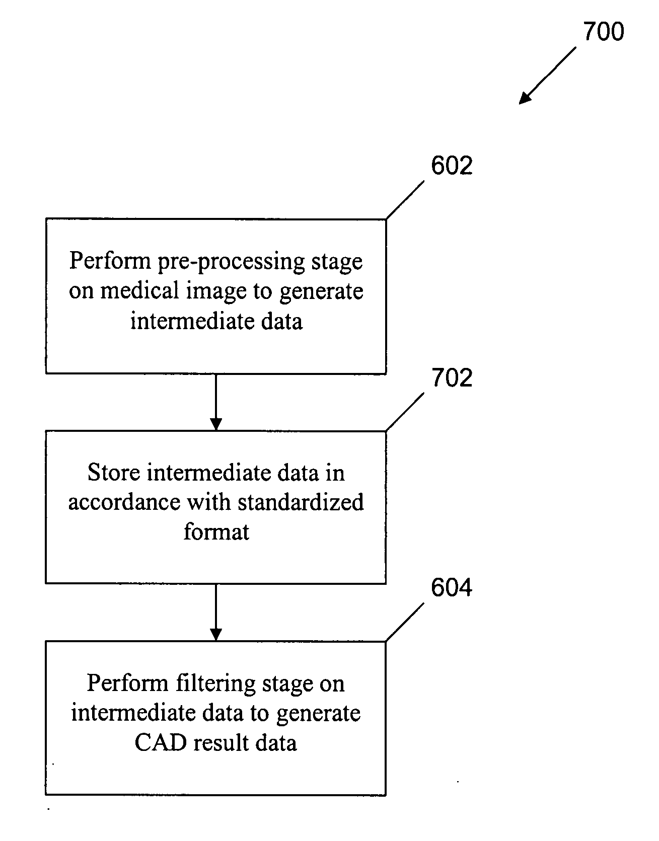
In a method of performing CAD analysis of a medical image, the medical image volume is processed in a pre-processing module to generate intermediate data, and the intermediate data is processed by a filtering module to generate CAD result data. The different modules may be executed by different processing nodes or devices, enabling a variety of different system architectures. The intermediate data may be stored in accordance with a standard, such as the Digital Imaging and Communications in Medicine (DICOM) standard. For instance, the intermediate data may be stored in accordance with the DICOM secondary capture image information object definition (SCI IOD) format. The CAD result data may be displayed on a viewer workstation, for example.

Description

CROSS-REFERENCE TO RELATED APPLICATION[0001]This application claims the benefit of the filing date of GB Patent Application No. 0604852.4, filed Mar. 9, 2006, which is incorporated herein by reference in its entirety.BACKGROUND OF THE INVENTION[0002]1. Field of the Invention[0003]The present invention relates to computer implemented medical image processing.[0004]2. Background Art[0005]Medical images are a valuable tool for diagnosis, and a variety of techniques or modalities have been developed, including but not limited to X-ray, computed tomography (CT), ultrasound, and magnetic resonance imaging (MRI). Conventionally, these images have been inspected visually by trained radiographers. The images may be generated digitally (CT and MRI) or digitized from an analog image (X-ray). The digitized images may be inspected and annotated by radiographers, and the annotations stored for later retrieval and analysis.[0006]The task of the radiographer can be made easier by storing the medica...

Claims


Application Information

Patent Timeline
IPC IPC(8): G06K9/00
CPCG06T7/0012
Inventor BEDDOE, GARETH RICHARD
Owner MEDICSIGHT
Why Eureka
Try Eureka