Systems and methods for providing diagnostic imaging studies to remote users

a diagnostic imaging and remote user technology, applied in the field of electronic distribution of diagnostic image studies to remote users, can solve the problems of reducing the efficiency of interpretation of images, occupying a large storage space, and difficult to transmit electronically from one location to another, so as to reduce the time and expense of sending

Inactive Publication Date: 2007-09-27
AGMEDNET
View PDF20 Cites 17 Cited by
  • Summary
  • Abstract
  • Description
  • Claims
  • Application Information

AI Technical Summary

Benefits of technology

[0016] In some embodiments, the compressed and encrypted images may be sent to one or more individuals that have a translator and a viewer. The translator these one or more individuals have may have hardware, software, or a combination of both that may decrypt and decompress the diagnostic imaging study and its components. This may allow individual to interpret the diagnostic image study by viewing them on the viewer. In some embodiments, the central server may cause the images to be sent to several individuals simultaneously, or substantially instantaneously, thus allowing for point to multipoint distribution of the images and thereby, reducing the time and expense for sending such images. In some embodiments, the encryption may be specific for a given individual.

Problems solved by technology

These images require a great deal of storage space and, therefore, may be difficult to transmit electronically from one location to another due to their size.
In addition, the size of these files increases the time required to transmit them and, thus, may tie up communication lines for extended periods of time and reduce the efficiency by which interpretations of the images may be obtained.
The JPEG algorithm is, however, “lossy,” meaning that the decompressed image that has been compressed by the algorithm isn't quite the same as the original.
As such, many DICOM web-based viewers using the JPEG algorithm may be unacceptable to an interpreter needing to provide conclusive diagnostic level interpretations.
Indeed, because the JPEG algorithm is lossy, an individual reviewing an image that has been compressed with the JPEG algorithm may not be able see important features of the image.
This loss of features may be unacceptable to those, for example, in the medical community when reviewing diagnostic imaging studies.
Lossless compression is naturally a more limited form of compression since it is necessary to preserve all information in the image.
As such, hospitals or other operators utilizing lossless compression may be limited in the number of vendors for imaging devices and PACS and, to have effective communication, all users of the system needing to examine / interpret the diagnostic imaging studies may be required to utilize the same hardware and / or proprietary compression / decompression software.
This may not be a problem in “closed” environments, but as soon as the need exists to transfer images to an individual outside the environment (for example, a doctor at another hospital), it may be difficult or impossible to effectively send lossless compressed image data to that individual.
These systems are limited, however, in the case of a remote user, defined as a user who is outside of the local area network.
The impact of this on the interpreting individual (for example, a radiologist or other doctor) is that if the images arrive fast, then they are of low resolution.
If they need to be of high resolution, then they will arrive much slower, or require all parties to share the same vendor system, thus limiting the number of physician relationships and access to specialists that a hospital or imaging center can have.
This severely slows the effective rate at which studies may be sent to an individual interpreter.
This problem may be exacerbated if the remote user is from a different hospital than where the PACS is located thus, making it more difficult to ensure that the remote user has a viewer that is compatible with a particular PACS.
In addition, these systems do not allow for the concurrent transfer of images.
Limiting the number of persons who may interpret images may severely reduce the profitability of a particular modality (imaging device) as well as access to qualified specialists for a particular patient.
In particular, the operator is not allowed to charge for a procedure until a final interpretation of the images has been completed.

Method used


Image

  • Systems and methods for providing diagnostic imaging studies to remote users
  • Systems and methods for providing diagnostic imaging studies to remote users
  • Systems and methods for providing diagnostic imaging studies to remote users

Examples


Embodiment Construction

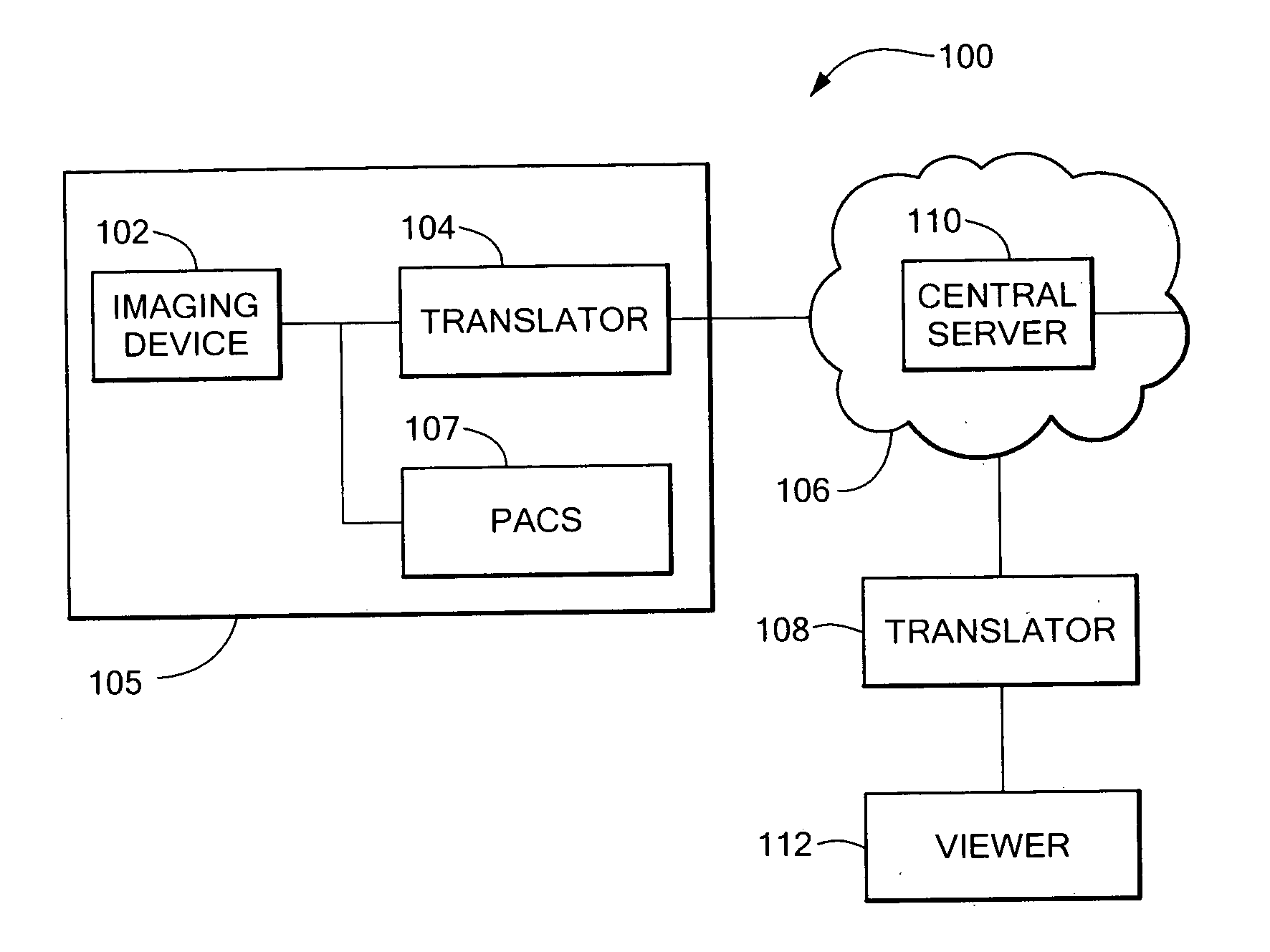
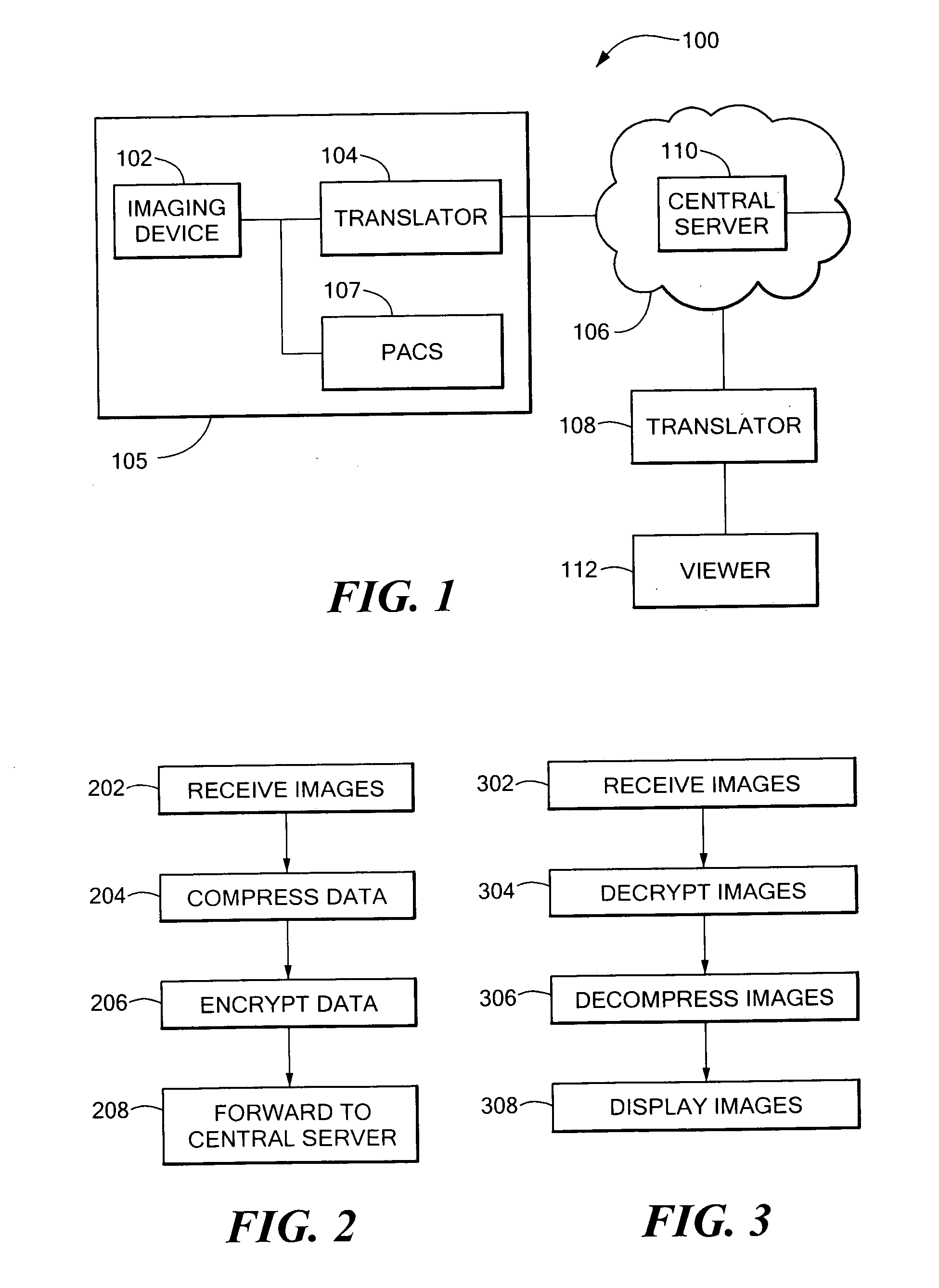
[0023] Referring now to FIG. 1, in some embodiments of the present invention, the system 100 may include imaging device 102. Of course, an imaging device is not required and the system may receive images from any source. This imaging device 102 may be any type of modality that may take diagnostic images of a patient. For example, the imaging device 102 may be an MRI machine, a digital X-ray machine, a CAT scan machine or any other type device. In other embodiments, the imaging device 102 may be any type of imaging device whether or not it is used for a diagnostic image of the human or not. For instance, the imaging device 102 could be an electron microscope or the like. The imaging device 102 transfers the images to other devices in the internal network 105 at the location where the imaging device 102 is located. For instance the internal network 105 could be a local area network (LAN) that has a plurality of devices connected to it. For instance, the imaging device 102 could be con...


PUM


Abstract

Systems and methods for the distribution of diagnostic imaging studies include a first translator electrically coupled to an imaging device, the first translator being arranged an configured to receive an diagnostic imaging study from the imaging device, compress and encrypt the diagnostic imaging study, and transfer the diagnostic imaging study to one or more additional translators substantially simultaneously. The system may also include a second translator, the second translator being arranged and configured to decrypt and decompress the diagnostic imaging study and a network, coupled between the first translator and the second translator, the network being arranged to transfer the diagnostic image study from the first translator to at least the second translator.

Description

CROSS REFERENCE TO RELATED APPLICATIONS [0001] This application is related to U.S. patent application Ser. No. ______, entitled Systems and Methods for Obtaining Readings of Diagnostic Imaging Studies, filed on even date herewith.TECHNICAL FIELD AND BACKGROUND ART [0002] The present invention relates to the transport and distribution of diagnostics image studies and, in particular, to the electronic distribution of diagnostic image studies to remote users. [0003] It is known in the prior art to utilize the Digital Imaging in Communication and Medicine (DICOM) standard to electronically transfer diagnostic imaging studies from a modality or PACS (Picture Archiving and Communications System) (also referred to herein as an imaging device) (both of which may be referred to as an “imaging device” herein) to a remote user. Examples of modalities include CAT scanners, X-ray machines, and MRI machines. The output of these devices may be referred to herein as a “diagnostic imaging study”. In...

Claims


Application Information

Patent Timeline
Patent Type & Authority Applications(United States)
IPC IPC(8): G06Q10/00G06K9/00G16H30/40G16H40/67
CPCG06Q50/22G06F19/321G16H40/67G16H30/40
Inventor GUTMAN, ABRAHAM
Owner AGMEDNET
Why Eureka
Try Eureka