Process and device for lung ventilation

a technology of lung ventilation and process, which is applied in the direction of valve operating means/release devices, applications, diagnostic recording/measuring, etc., can solve problems such as hospital complications, and achieve the effect of improving the ventilation of the lungs

Active Publication Date: 2007-10-25
DRAGERWERK AG
View PDF0 Cites 25 Cited by
  • Summary
  • Abstract
  • Description
  • Claims
  • Application Information

AI Technical Summary

Benefits of technology

[0014]A special advantage of the present invention is the possibility of gently respirating the patient with improved ventilation of the lungs during and after a medical and especially surgical procedure with possible development of atelectasis.
[0015]An exemplary embodiment of the present invention will be explained below by means of the only figure, which schematically shows the principal components of a device for carrying out the p

Problems solved by technology

Even though the administration of oxygen and breathing training can be employed highly efficiently in most cases, respiratory failure may develop during the early postoper

Method used


Image

  • Process and device for lung ventilation
  • Process and device for lung ventilation

Examples


Embodiment Construction

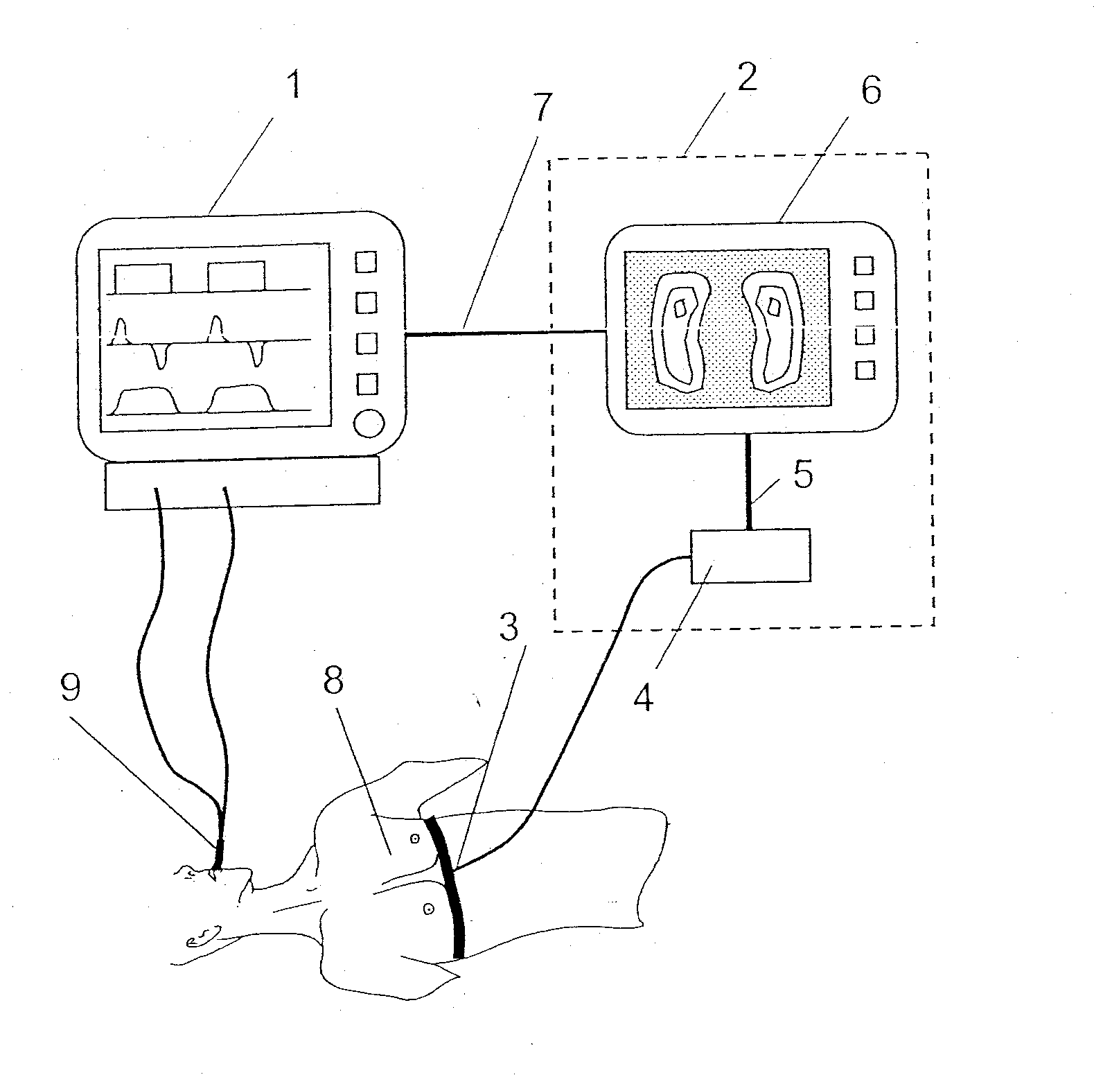
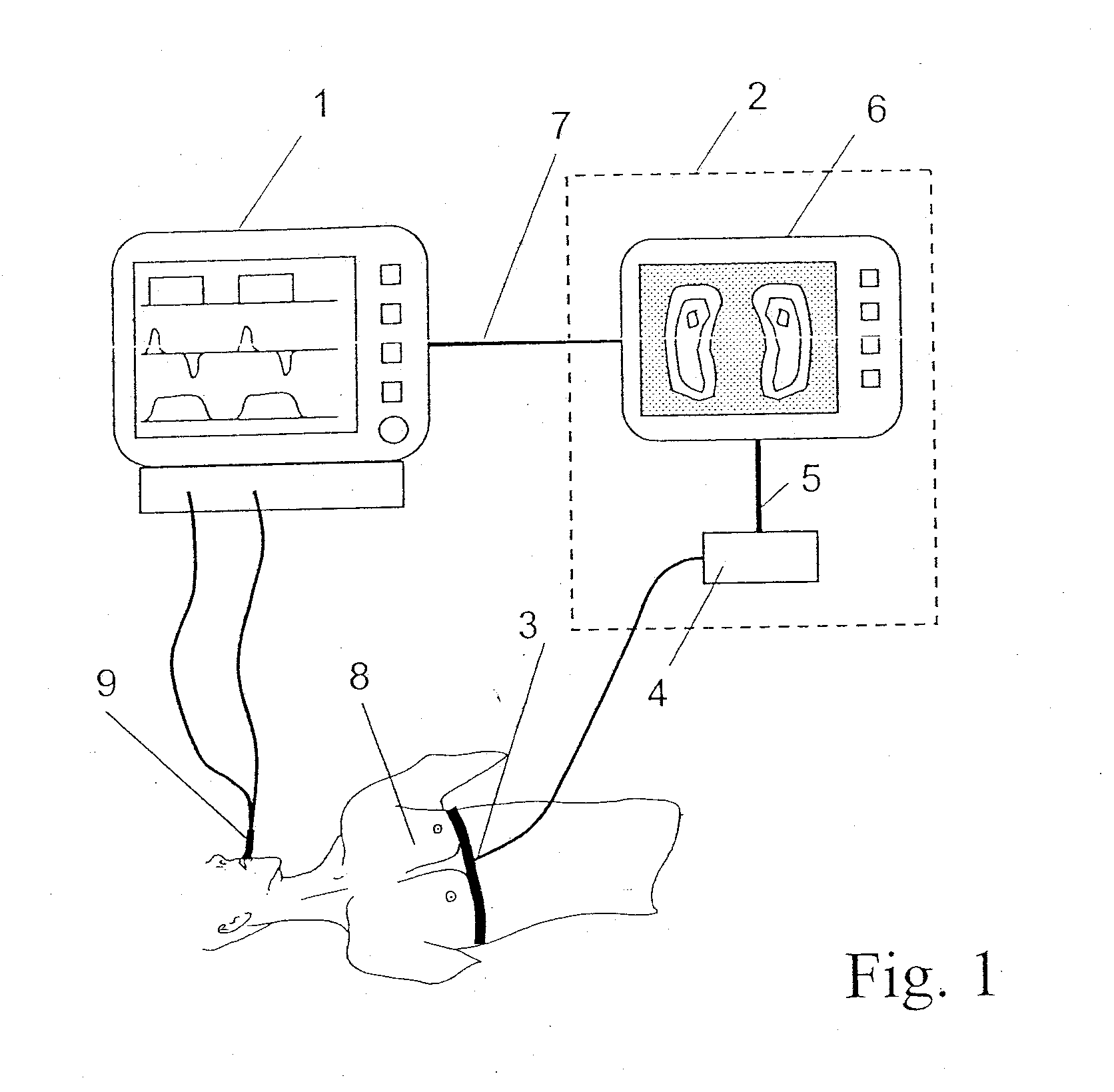
[0018]Referring to the drawing in particular, a first image of the lungs, which are not yet compromised by the anesthesia, is determined by means of the EIT system 2 for the patient 8, in the recumbent position, before initiation of anesthesia. The patient 8 will have to undergo, for example, major surgery.

[0019]The EIT system 2 contains, as is shown, an evaluating and display unit 6 and a computing unit 4. The system 2 detects images of the lung status of the patient 8 with the use of an electrode belt 3.

[0020]The computing unit 4 may also be arranged separately outside the EIT system 2, for example, between the evaluating and display unit 6 and the respirator 1. The computing unit 4 is connected to the evaluating and display unit 6 by means of the line 5. Furthermore, a technical alternative is that the EIT system 2 is arranged in an integrated total system together with the computing unit 4 and the respirator 1. While the electrodes remain in the same position as before, a second...


PUM


Abstract

A process and a device is presented with an electro-impedance tomography (EIT) system (2), with a computing unit (4) and with a respirator (1) for gentle mechanical lung ventilation especially in case of atelectases following surgical procedures. A first image of the healthy lungs is first recorded prior to anesthetization by means of the EIT system (2) and the total area and/or the spatial distribution of ventilated lung areas are determined from the image values by means of the computing unit (4). Second or additional status images are recorded after assumed lung damage and the total area and/or the spatial distribution of ventilated lung areas are determined. The total areas and/or the spatial distribution of the ventilated lung areas from the first and second or further status images are compared by means of the computing unit (4) and analyzed for the presence of lung areas that have no or reduced ventilation due to atelectases. The information on the presence, the extent and/or the spatial distribution of atelectases is sent by the EIT system (2) to the respirator (1) so that the respiration pressure is increased step by step by the respirator (1) as a function of the status image or the currently determined status images until the current image of the lung status corresponds to the first status image or comes close to it with minimal deviations. The respiration pressure is subsequently reduced again step by step by the respirator (1) until the computing unit (4) detects a reduction of the ventilated lung areas and the respiration pressure is subsequently increased again by means of the respirator (1) to the last value at which no change occurred in the ventilated lung areas.

Description

CROSS REFERENCE TO RELATED APPLICATIONS[0001]This application claims the benefit of priority under 35 U.S.C. §119 of German Patent Application DE 10 2006 018 198.0 filed Apr. 19, 2006, the entire contents of which are incorporated herein by reference.FIELD OF THE INVENTION[0002]The present invention pertains to a process and a device for lung ventilation (also known as lung respiration).BACKGROUND OF THE INVENTION[0003]Electric impedance tomography (EIT) is a process, which is known per se, in which the electric impedance between the feed point and the test point can be calculated by feeding an alternating electric current of, e.g., 5 mAcff at 50 kHz into any electrically conductive body, here preferably into the human body, and measuring the surface potentials resulting therefrom at different points of the body. A two-dimensional tomogram of the electric impedance distribution in the body being considered can be determined on the basis of suitable mathematical reconstruction algori...

Claims


Application Information

Patent Timeline
IPC IPC(8): A62B7/00
CPCA61B5/0536A61M16/00A61M2205/3569A61M2205/50A61M2230/65A61M2230/005A61M16/022
Inventor TESCHNER, ECKHARDWEISMANN, DIETER
Owner DRAGERWERK AG
Why Eureka
Try Eureka