

Compositions and methods for detecting keratoconus
a technology of keratoconus and composition, applied in the field of materials and methods for detecting keratoconus, can solve the problem of remaining undetectabl
- Summary
- Abstract
- Description
- Claims
- Application Information
AI Technical Summary
Benefits of technology
Problems solved by technology
Method used
Image

Examples
example 1
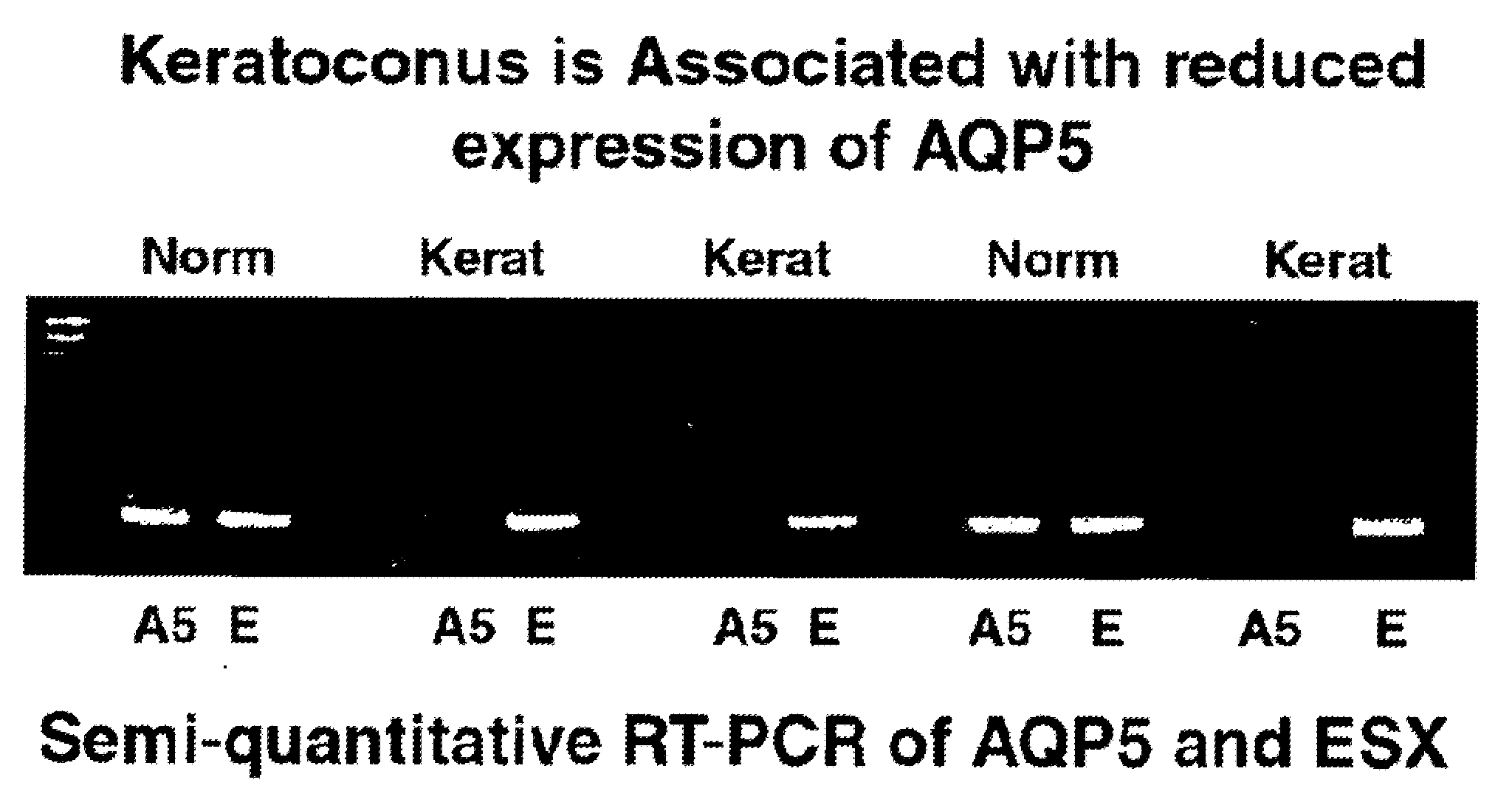
Aquaporin 5: A Molecular Marker for Keratoconus
[0081]This example demonstrates the suppression of Aquaporin 5 (AQP5) in keratoconus (KC) corneas transplant buttons. In this experiment, both the surgeon and the molecular geneticist performing the molecular work knew the diagnosis of the specimens submitted for RT-PCR analysis.
[0082]i) Tissue Procurement
[0083]a) cDNA Library
[0084]Seven KC corneal host buttons approximately 7.5 mm in size were removed at the time of penetrating keratoplasty. A piece of tissue was sent for histopathology, and the rest immediately immersed in preservative (RNAlater, Ambion, Austin Tex.). All patients had moderate to advanced KC, with some having central corneal scarring. All patients had steep keratometry readings in excess of 60D and were contact lens intolerant. None of the patients had worn contact lenses for 3 months prior to undergoing corneal transplantation.
[0085]b) RT-PCR Experiments on Whole Corneas
[0086]Two additional corneal buttons were remov...
example 2
Aquaporin 5: A Molecular Marker For Detecting Sub Clinical Disease
[0096]This example demonstrates the suppression of Aquaporin 5 (AQP5) in keratoconus (KC) corneal transplant buttons, and use of this information to design a molecular genetic test for confirming the presence of sub clinical KC.
[0097]Material and Methods
[0098]Study Specimens and Tissue Procurement
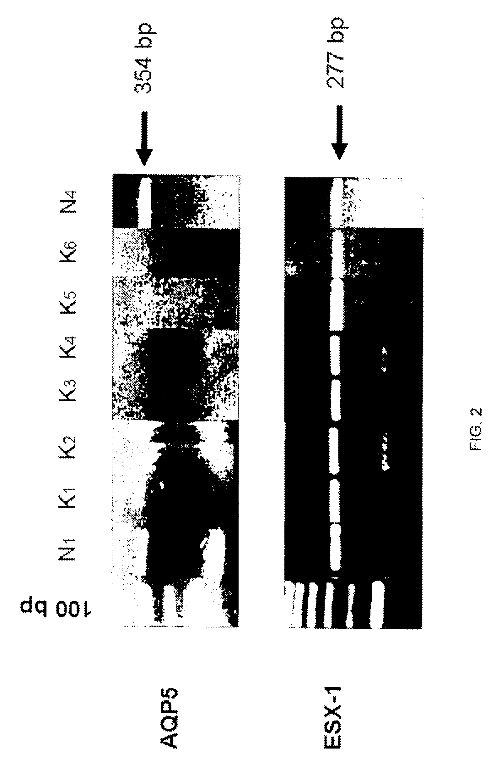
[0099]Seven advanced KC(K readings greater than 60D) corneal buttons approximately 7.5 mm in diameter and two non KC buttons(one with trauma and one with Fuchs dystrophy) were immersed immediately in preservative (RNAlater, Ambion, Austin, Tex.) at the time of keratoplasty. 3 donor corneal scleral rim specimens were obtained from normal donor corneas and, one 7 mm diameter specimen of epithelium was also procured from a normal 6D myope at the time of PRK.
[0100]To test whether corneal epithelial samples alone would be adequate for diagnostic purposes we also compared three 9 mm epithelial specimens from myopic patients at the ...
examples 1 and 2
[0112]1. Amsler M. Le keratoconus fruste au javal. Ophthalmologica 1938; 96:77-83.[0113]2. Li X, Rabinowitz Y S, Rasheed K, Yang H. Longitudinal study of the normal eyes in unilateral keratoconus. Ophthalmology 2004; 111:44046.[0114]3. Binder P S, Lindstrom R, Stulting R D, Donnenfeld E, Wu H, McDonnell P, Rabinowitz Y S, Keratoconus and corneal ectasia after LASIK. J Refract Surg 2005; 21:749-52[0115]4. Randleman J B, Russell B, Ward M A, Thompson K P, Stulting R D. Risk factors and prognosis for corneal ectasia after LASIK. Ophthalmology 2003; 110:267-75.[0116]5. Ambrosio R Jr, Klyce S D, Wilson S E. Corneal topographic and pachymetric screening of keratorefractive patients. J Refract Surg 2003; 19:24-29[0117]6. Lifshitz T, Levy J, Klemperer I, Levinger S. Late bilateral keratectasia after LASIK in a low myopic patient. J Refract Surg. September-October 2005;21(5):494-6.[0118]7. Agre P, Kozono D. Aquaporin water channels: molecular mechanisms for human diseases. FEBS Lett 2003: 55...
PUM
| Property | Measurement | Unit |
|---|---|---|
| Fraction | aaaaa | aaaaa |
| Substance count | aaaaa | aaaaa |
| Atomic weight | aaaaa | aaaaa |
Abstract
Description
Claims
Application Information