Implantable Medical Device with Electromechanical Delay Measurement for Lead Position and Ventricular

a technology of lead position and ventricular, applied in the field of implantable medical devices and cardiac rhythm management systems, can solve the problems of poor quality of life, easy tire, and compromise of cardiac therapy or monitoring benefits

Active Publication Date: 2007-11-01
MEDTRONIC INC
View PDF24 Cites 35 Cited by
  • Summary
  • Abstract
  • Description
  • Claims
  • Application Information

AI Technical Summary

Problems solved by technology

Dislodgement or migration of leads can compromise the benefits of cardiac therapy or monitoring by shifting the location of the electrodes (as well as any physiologic s

Method used


Image

  • Implantable Medical Device with Electromechanical Delay Measurement for Lead Position and Ventricular
  • Implantable Medical Device with Electromechanical Delay Measurement for Lead Position and Ventricular
  • Implantable Medical Device with Electromechanical Delay Measurement for Lead Position and Ventricular

Examples


Embodiment Construction

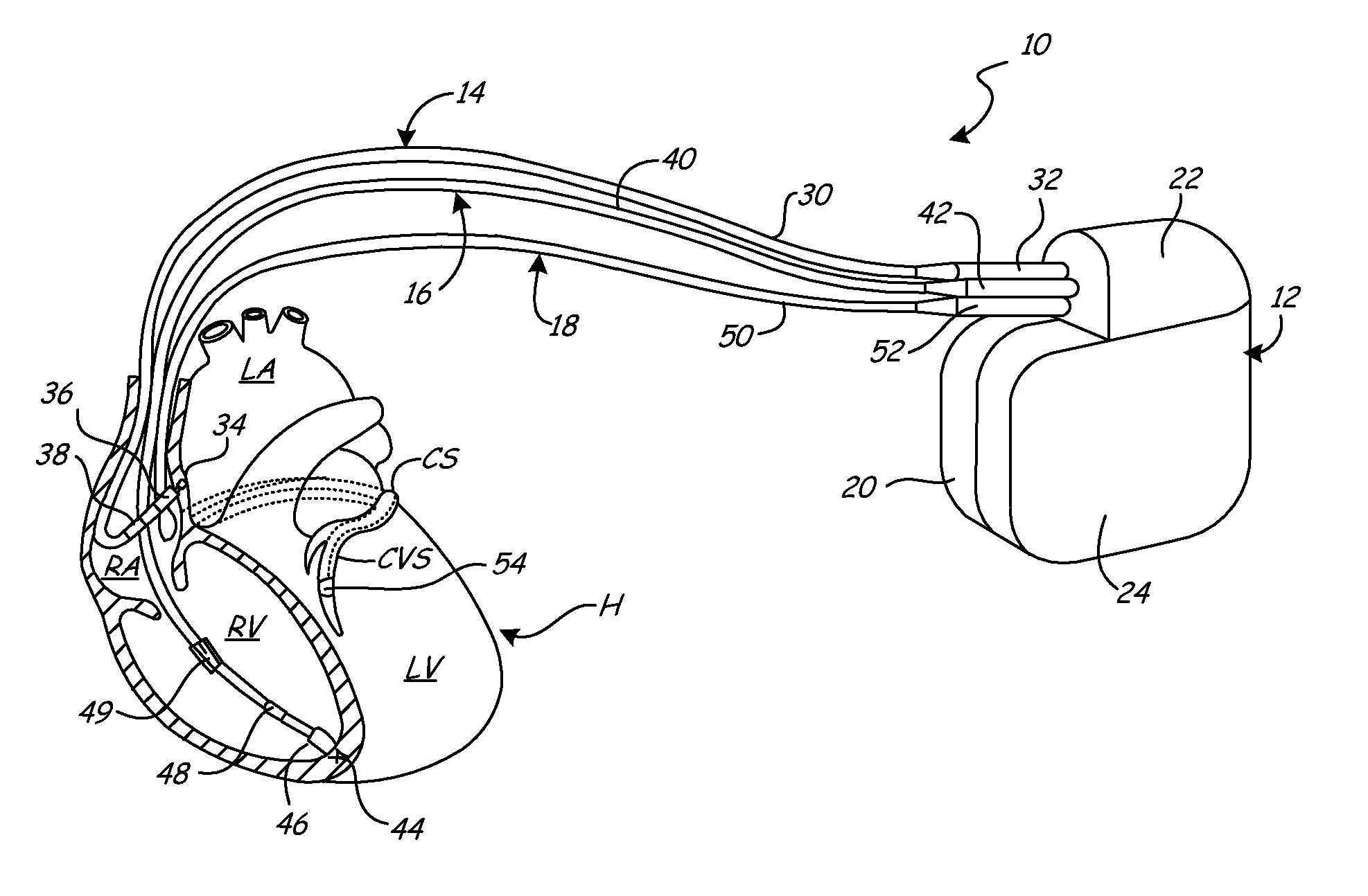
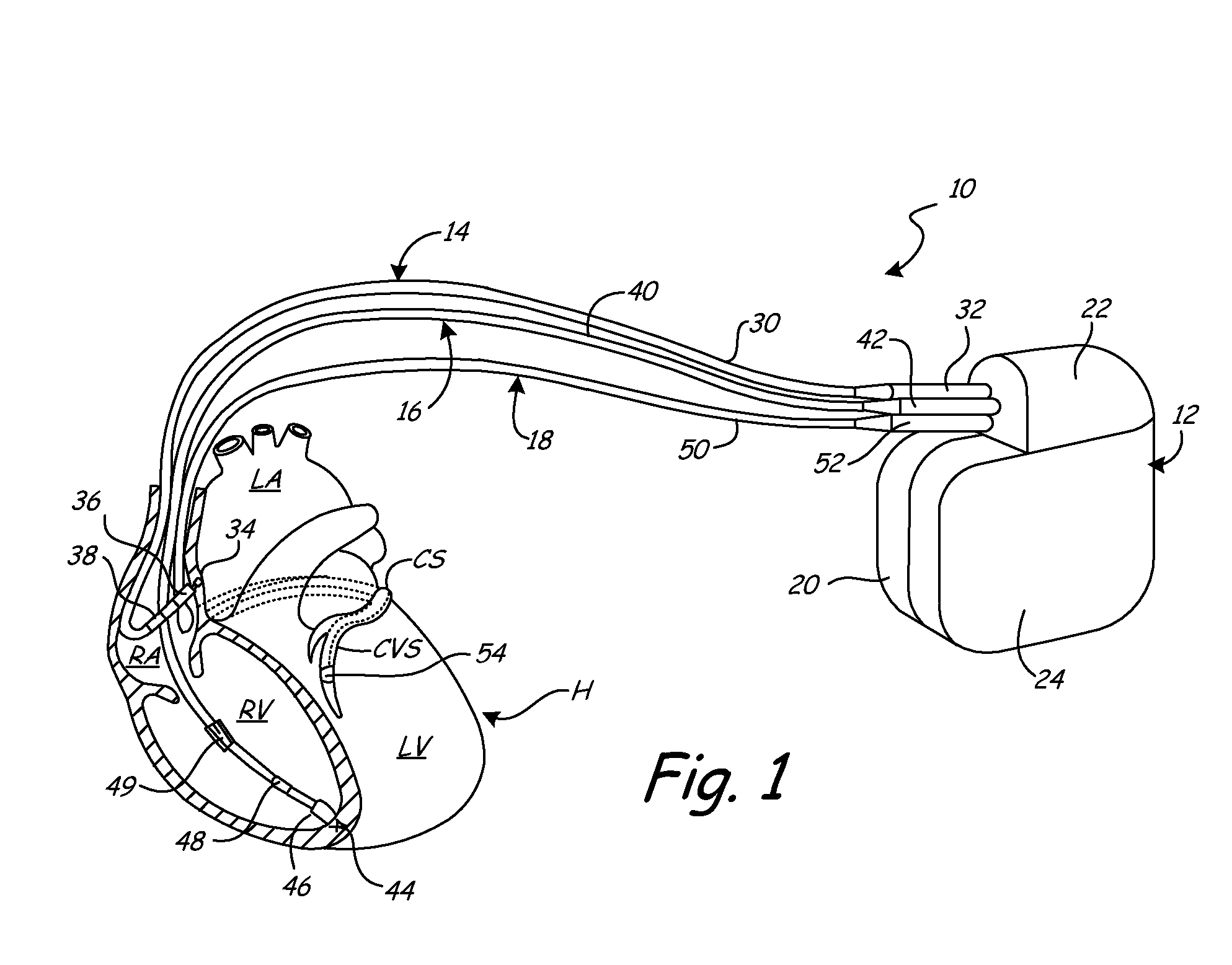
[0015]FIG. 1 shows cardiac resynchronization therapy (CRT) system 10, which restores ventricular synchronization in heart H by delivering pacing pulses to one or more chambers of heart H. In FIG. 1, heart H is shown in a partially cutaway view illustrating right atrium RA, left atrium LA, right ventricle RV, left ventricle LV, coronary sinus CS, and coronary venous system CVS.

[0016] CRT system 10 includes implantable pulse generator (IPG) 12, right atrial (RA) lead 14, right ventricular (RV) lead 16, and left ventricular (LV) lead 18. As shown in FIG. 1, IPG 12 includes housing or canister 20, header 22 and can electrode 24. The circuitry and power source of IPG 12 are located within housing 20. The circuitry communicates with leads 14, 16, and 18 through electrical connectors within header 22. Can electrode 24 is formed on or is a part of the outer surface of housing 20, and acts as a remote indifferent electrode with respect to one or more of the electrodes carried by leads 14, 1...


PUM


Abstract

The cardiac rhythm management system includes an implantable medical device (IMD) with leads carrying electrodes for sensing cardiac electrical activity, and a physiologic sensor for sensing cardiac mechanical activity. The IMD measures electromechanical delays between electrical activity sensed by the electrodes and mechanical activity sensed by the physiologic sensor. The measured electromechanical delays can be used to detect lead dislodgement and to assess dyssynchrony between two areas of the heart, such as the right ventricle and the left ventricle.

Description

BACKGROUND OF THE INVENTION [0001] The present invention relates generally to implantable medical devices (IMDs) and cardiac rhythm management systems. In particular, the present invention relates to measurement of electromechanical delays, which can be used to detect lead displacement or migration of electrical leads, or to assess ventricular dyssynchrony. [0002] IMDs, such as pacemakers, implantable cardioverter defibrillators and implantable hemodynamic monitors, are used to monitor cardiac conditions and deliver therapy when appropriate. The IMDs typically include electrical leads that carry electrodes for pacing, cardioverting, defibrillating, sensing and monitoring functions. The leads may also include a physiologic sensor, such as a pressure sensor, oxygen sensor, temperature sensor, flow sensor, or accelerometer. [0003] Dislodgement or migration of leads can compromise the benefits of cardiac therapy or monitoring by shifting the location of the electrodes (as well as any ph...

Claims


Application Information

Patent Timeline
IPC IPC(8): A61N1/00
CPCA61N1/3622A61N2001/083A61N1/3627
Inventor CHO, YONG K.GEIMAN FERRI, BETH J.BENNETT, TOMMY D.
Owner MEDTRONIC INC
Why Eureka
Try Eureka