Methods for Culturing Keratinocytes from Human Embryonic Stem Cells

a technology of embryonic stem cells and keratinocytes, which is applied in the field of isolating and culturing human keratinocytes from embryonic stem cells, can solve the problems of deteriorating growth potential of such colonies, medium has serious limitations for the growth of es-cell-derived keratinocytes, and culture systems previously developed for growing post-natally derived keratinocytes are not optimal for the continued growth of es-cell

Inactive Publication Date: 2007-11-29
PRESIDENT & FELLOWS OF HARVARD COLLEGE
View PDF0 Cites 7 Cited by
  • Summary
  • Abstract
  • Description
  • Claims
  • Application Information

AI Technical Summary

Benefits of technology

[0012] In addition, we have developed methods of producing a mega-embryoid body that is useful for obtaining embryonic stem cells. The mega-embryoid bodies prepared using the methods of the invention are larger than standard embryoid bodies prepared using conventional methods. In addition the mega-embryoid bodies allow faster harvesting of keratinocytes than does use of standard embryoid bodies in keratinocyte harvest methods.

Problems solved by technology

Several issues arise in this connection: Because ES cells are capable of forming teratomas, it is important to free these cell types from all remaining ES cells before their use.
But on serial transfer, the growth potential of such colonies deteriorates.
This seems to mean that the culture systems that have previously developed for growing post-natally derived keratinocytes are not optimal for the continuing growth of ES-cell derived keratinocytes.
We have also found that this medium has the serious limitation for the growth of ES cell-derived keratinocytes that the cells will not tolerate dilution in it.

Method used


Image

  • Methods for Culturing Keratinocytes from Human Embryonic Stem Cells
  • Methods for Culturing Keratinocytes from Human Embryonic Stem Cells
  • Methods for Culturing Keratinocytes from Human Embryonic Stem Cells

Examples


example 1

Introduction

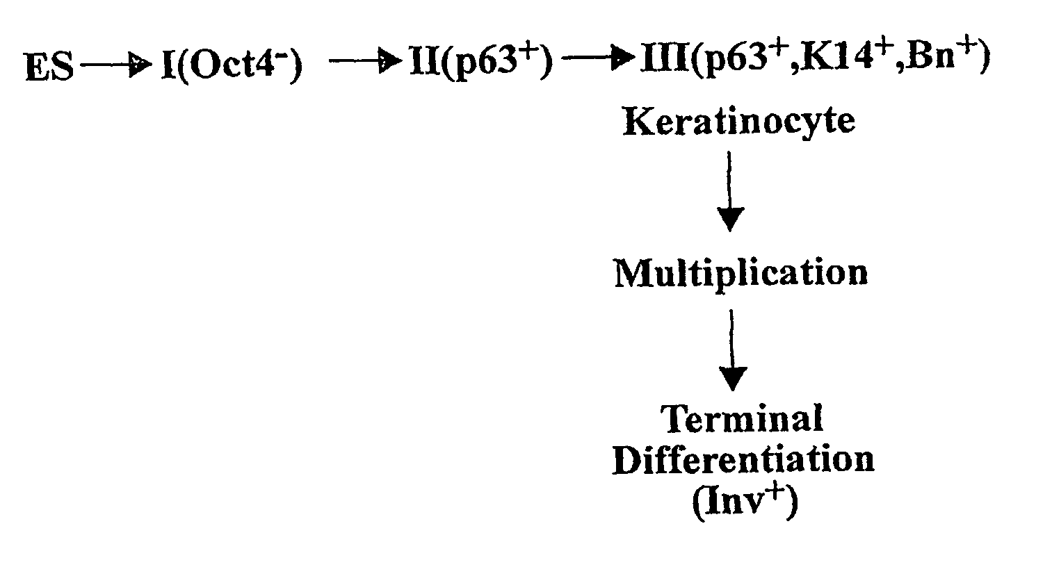
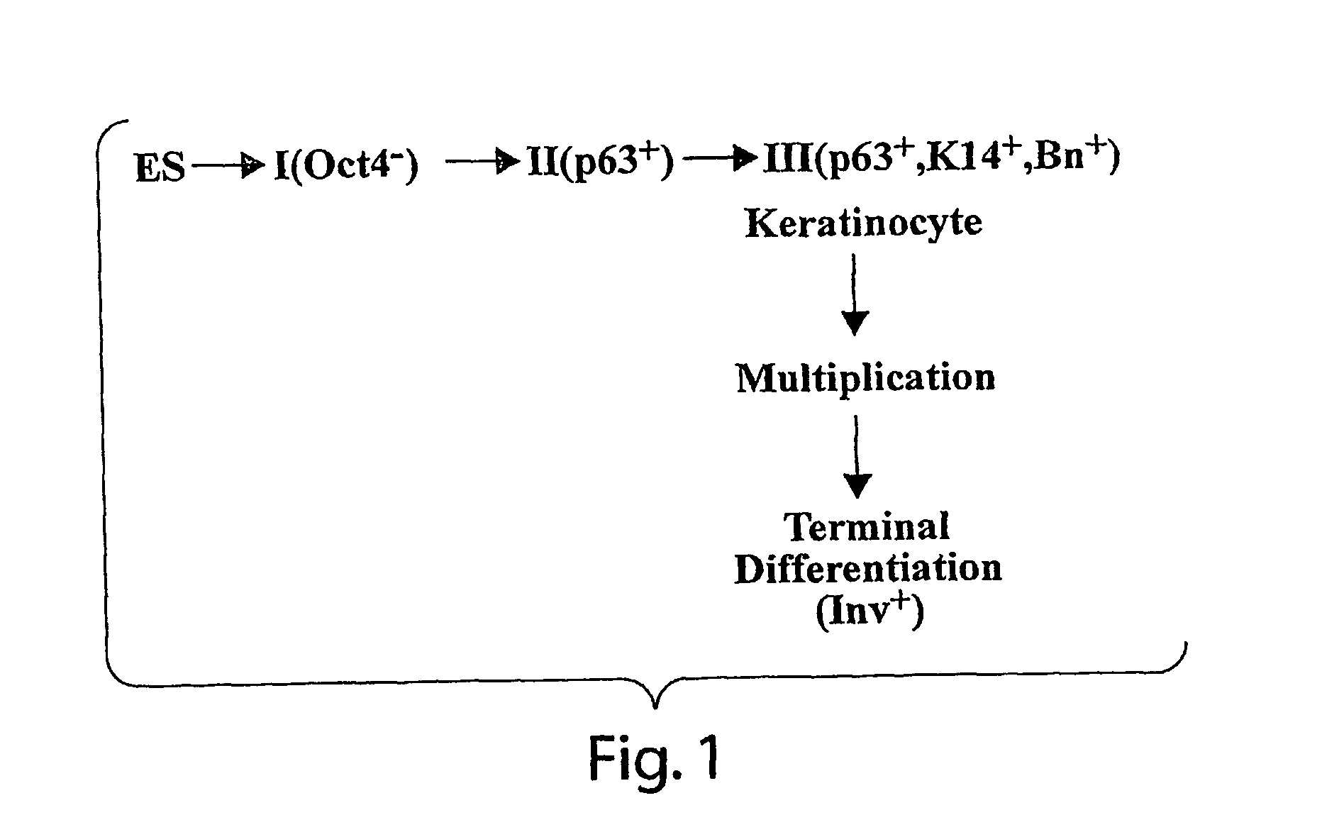
[0067] Human embryonic stem cells injected into scid mice produce nodules containing differentiated somatic tissues. From the trypsinized cells of such a nodule, we have recovered keratinocytes that can be grown in cell culture. The method of recovery is sensitive enough to detect small numbers of keratinocytes formed in the nodule, but for purposes of analysis, it is preferable to study the development of the entire keratinocyte lineage in culture. The principle of our analysis is the successive appearance of markers, including transcription factors with considerable specificity for the keratinocyte (p63 and basonuclin) and differentiation markers characteristic of its final state (keratin 14 and involucrin). We have determined the order of marker succession during the time- and migration-dependent development of keratinocytes from single embryoid bodies in cell culture. Of the markers we have examined, p63 was the earliest to appear in the keratinocyte lineage. The s...

example 2

Background

[0094] Much of the general enthusiasm for research on human embryonic stem cells is based on the possible therapeutic use of derived somatic cell types. Several issues arise in this connection: Because ES cells are capable of forming teratomas, it is important to free these cell types from all remaining ES cells before their use. To achieve purity, it is important that the derived somatic cell type of interest be made serially cultivable, so it can be clonally isolated. There are currently no examples cited in the literature for any cell type derived from human ES cells.

[0095] In addition, the somatic cell types derived from ES cells must be examined to determine whether they are identical to the similar somatic type isolated from post-natal or fetal tissues. ES cells are a cultured cell type, not an implanted blastocyst; therefore, clues that establish order in embryos (such as polarity and gradients) are absent. If important cues are missing, the cell types generated ...

example 3

Mega Embryoid Bodies (EBs) and their Conversion to Keratinocytes

[0102] We have developed a new method of preparing EBs so that they are much larger than those prepared by conventional methods. We call them Mega EBs or multilocular EBs. They are produced in a flask whose geometry favors larger scale aggregation on the bottom. We have developed conditions permitting EBs so produced, when transferred to tissue culture dishes, to attach very quickly and cell migration onto the surface of the dish begins almost immediately. Differentiation along the keratinocyte lineage occurs much more rapidly than in earlier experiments. On the very day following introduction of an EB to the tissue culture dish, p63-containing cells appear in the zone of migration and a great many p63-containing cells appear by five days. This suggests that keratinocyte formation from cultured EBs is greatly accelerated in comparison with normal human embryogenesis. In the mouse, p63 appears at between ⅓ and ½ of the...


PUM

PropertyMeasurementUnit
timeaaaaaaaaaa
timeaaaaaaaaaa
lengthaaaaaaaaaa

Abstract

The invention relates to methods for isolating and culturing human keratinocytes from embryonic stem cells. The methods are useful for producing substantially pure cultures of keratinocytes.

Description

RELATED APPLICATION [0001] This application claims priority under 35 U.S.C. § 119 from U.S. provisional application Ser. No. 60 / 527,920, filed Dec. 8, 2003, the contents of which is incorporated herein in its entirety.GOVERNMENT SUPPORT [0002] This invention was made in part with government support under grant number R01GM068478-01 from the National Institutes of Health (NIH). The government may have certain rights in this invention.FIELD OF THE INVENTION [0003] The invention relates to methods of isolating and culturing human keratinocytes from embryonic stem cells. The methods are useful for producing substantially pure cultures of keratinocytes. BACKGROUND OF THE INVENTION [0004] The ability to culture keratinocytes has enabled advancements in the treatment of burns and trauma that affect the skin. The likelihood of survival of patients with severe burns has improved dramatically with the advent of methods to replace lost skin with laboratory-grown skin cells. One source of repla...

Claims


Application Information

Patent Timeline
Patent Type & Authority Applications(United States)
IPC IPC(8): C12N5/06C12N5/08C12N5/071
CPCA61K35/12C12N5/0629C12N2500/14C12N2502/13C12N2501/385C12N2501/70C12N2506/02C12N2500/99C12N2500/90
Inventor GREEN, HOWARDIUCHI, SHIROEASLEY, KAREN
Owner PRESIDENT & FELLOWS OF HARVARD COLLEGE
Why Eureka
Try Eureka