Methods and systems for treating tumors using electroporation

a tumor and electroporation technology, applied in the field of electroporation, can solve problems such as inability to closely monitor and control

Inactive Publication Date: 2008-01-17
ANGIODYNAMICS INC
View PDF99 Cites 98 Cited by
  • Summary
  • Abstract
  • Description
  • Claims
  • Application Information

AI Technical Summary

Benefits of technology

[0020] Accordingly, an object of the present invention is to provide improved systems and methods for treating tumor sites using electroporation.
[0021] Another object of the present invention is to provide systems and method for treating tumor sites using electroporation using sufficient electrical pulses to induce electroporation of cells in the tumor tissue site, without creating a thermal damage effect to a majority of the tumor tissue site.
[0026] Yet another object of the present invention is to provide systems and methods for treating tumor sites using electroporation that is performed in a controlled manner, with a proper selection of voltage application time.
[0028] Still a further object of the present invention is to provide systems and methods for treating tumor sites using electroporation that is performed in a controlled manner to provide for controlled pore formation in cell membranes.
[0029] Still another object of the present invention is to provide systems and methods for treating tumor sites using electroporation that is performed in a controlled manner to create a tissue effect in the cells at the tumor tissue site while preserving surrounding tissue.

Problems solved by technology

However, when used deep in the body, as opposed to the outer surface or in the vicinity of the outer surface of the body, it has a drawback that is typical to all minimally invasive surgical techniques that occur deep in the body, it cannot be closely monitored and controlled.

Method used


Image

  • Methods and systems for treating tumors using electroporation
  • Methods and systems for treating tumors using electroporation
  • Methods and systems for treating tumors using electroporation

Examples


example 2

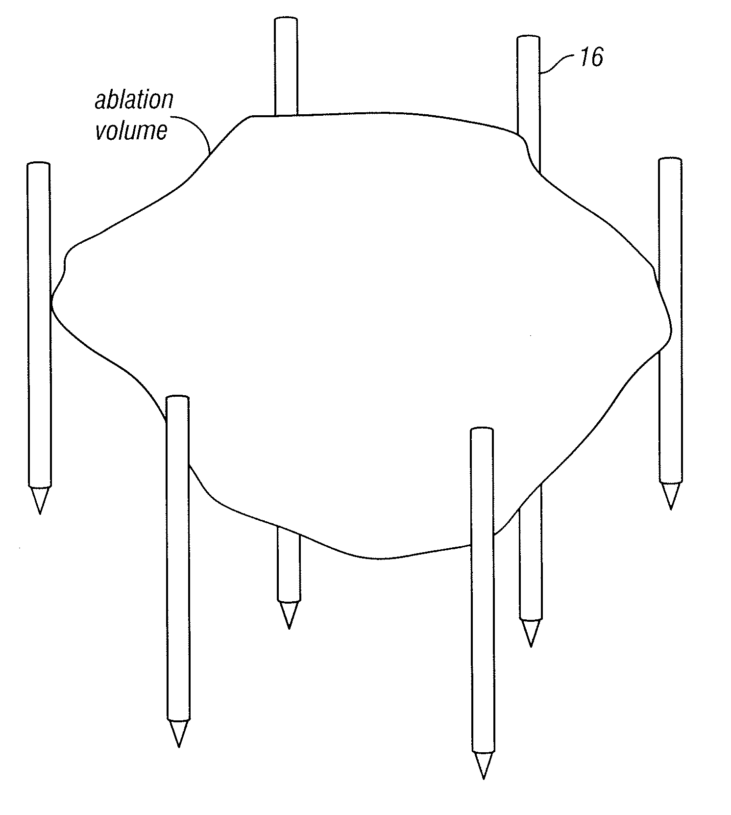
[0081] An area of the lung tumor tissue site is imaged. The array 16 of electrodes is introduced to the lung tumor tissue site, and positioned in a surrounding relationship to the lung tumor tissue site. Imaging is used to confirm that the electrodes are properly placed. The two electrodes are separated by a distance of 5 mm to 10 cm at various locations of the lung tumor tissue site. Pulses are applied with a duration of about 90 to 110 microseconds each. Monitoring is performed using a CT scan. The lung tumor tissue site is monitored. In response to the monitoring, pulses are adjusted to maintain a temperature of no more than 75 degrees C. A voltage gradient at the lung tumor tissue site in a range of from about 50 volt / cm to about 5000 volt / cm is created. A volume of the lung tumor tissue site undergoes cell necrosis.

example 3

[0082] An area of the breast tumor tissue site is imaged. The array 16 of electrodes is introduced to the breast tumor tissue site, and positioned in a surrounding relationship to the breast tumor tissue site. Imaging is used to confirm that the electrodes are properly placed. Pulses are applied with a duration of about 100 microseconds each. A monitoring electrode 18 is utilized. Prior to the full electroporation pulse being delivered a test pulse is delivered that is about 10% of the proposed full electroporation pulse. The test pulse does not cause irreversible electroporation. The breast tumor tissue site is monitored. In response to the monitoring, pulses are adjusted to maintain a temperature of no more than 60 degrees C. A voltage gradient at the breast tumor tissue site in a range of from about 50 volt / cm to about 8000 volt / cm is created. A volume of the breast tumor tissue site of undergoes cell necrosis.

example 4

[0083] An area of the brain tumor tissue site is imaged. A array 16 of electrodes is introduced to the brain tumor tissue site, and positioned in a surrounding relationship to the brain tumor tissue site. Imaging is used to confirm that the array 16 of electrodes is properly placed. Pulses are applied with a duration of 5 microseconds to about 62 seconds each. Monitoring is preformed using ultrasound. The brain tumor tissue site is monitored. In response to the monitoring, pulses are adjusted to maintain a temperature of no more than 100 degrees C. A voltage gradient at the brain tumor tissue site in a range of from about 50 volt / cm to about 1000 volt / cm is created. A volume of the brain tumor tissue site undergoes cell necrosis.


PUM


Abstract

A system is provided for treating tumor tissue sites of a patient. At least first and second mono-polar electrodes are configured to be introduced at or near the tumor tissue site of the patient. A voltage pulse generator is coupled to the first and second mono-polar electrodes. The voltage pulse generator is configured to apply sufficient electrical pulses between the first and second mono-polar electrodes to induce electroporation of cells in the tumor tissue site, to create necrosis of cells of the tumor tissue site, but insufficient to create a thermal damaging effect to a majority of the tumor tissue site.

Description

CROSS-REFERENCE TO RELATED APPLICATIONS [0001] This application a divisional of U.S. Ser. No. 11 / 165,961, filed Jun. 24, 2005, and is related to U.S. Ser. Nos. 11 / 165,881 filed Jun. 24, 2005 and 11 / 165,908, filed Jun. 24, 2005, all of which applications are fully incorporated herein by reference.BACKGROUND [0002] 1. Field of the Invention [0003] This invention relates generally to electroporation, and more particularly to systems and methods for treating tumor tissue sites of a patient using electroporation. [0004] 2. Description of the Related Art [0005] Electroporation is defined as the phenomenon that makes cell membranes permeable by exposing them to certain electric pulses (Weaver, J. C. and Y. A. Chizmadzhev, Theory of electroporation: a review. Bioelectrochem. Bioenerg., 1996. 41: p. 135-60). The permeabilization of the membrane can be reversible or irreversible as a function of the electrical parameters used. In reversible electroporation the cell membrane reseals a certain ...

Claims


Application Information

Patent Timeline
Patent Type & Authority Applications(United States)
IPC IPC(8): A61B18/00
CPCA61B18/1477A61B2018/0016A61B2018/143A61B2018/00797A61B2018/00577A61B2018/00613
Inventor RUBINSKY, BORISONIK, GARYMIKUS, PAUL
Owner ANGIODYNAMICS INC
Why Eureka
Try Eureka