
Methods and Apparatus for Volume Computer Assisted Reading Management and Review

a volume computer and reading technology, applied in image analysis, medical science, image enhancement, etc., can solve the problems of enormous number of ct slices and tedious and time-consuming tasks

Inactive Publication Date: 2008-01-24
GENERAL ELECTRIC CO
View PDF22 Cites 39 Cited by
  • Summary
  • Abstract
  • Description
  • Claims
  • Application Information

AI Technical Summary

Benefits of technology

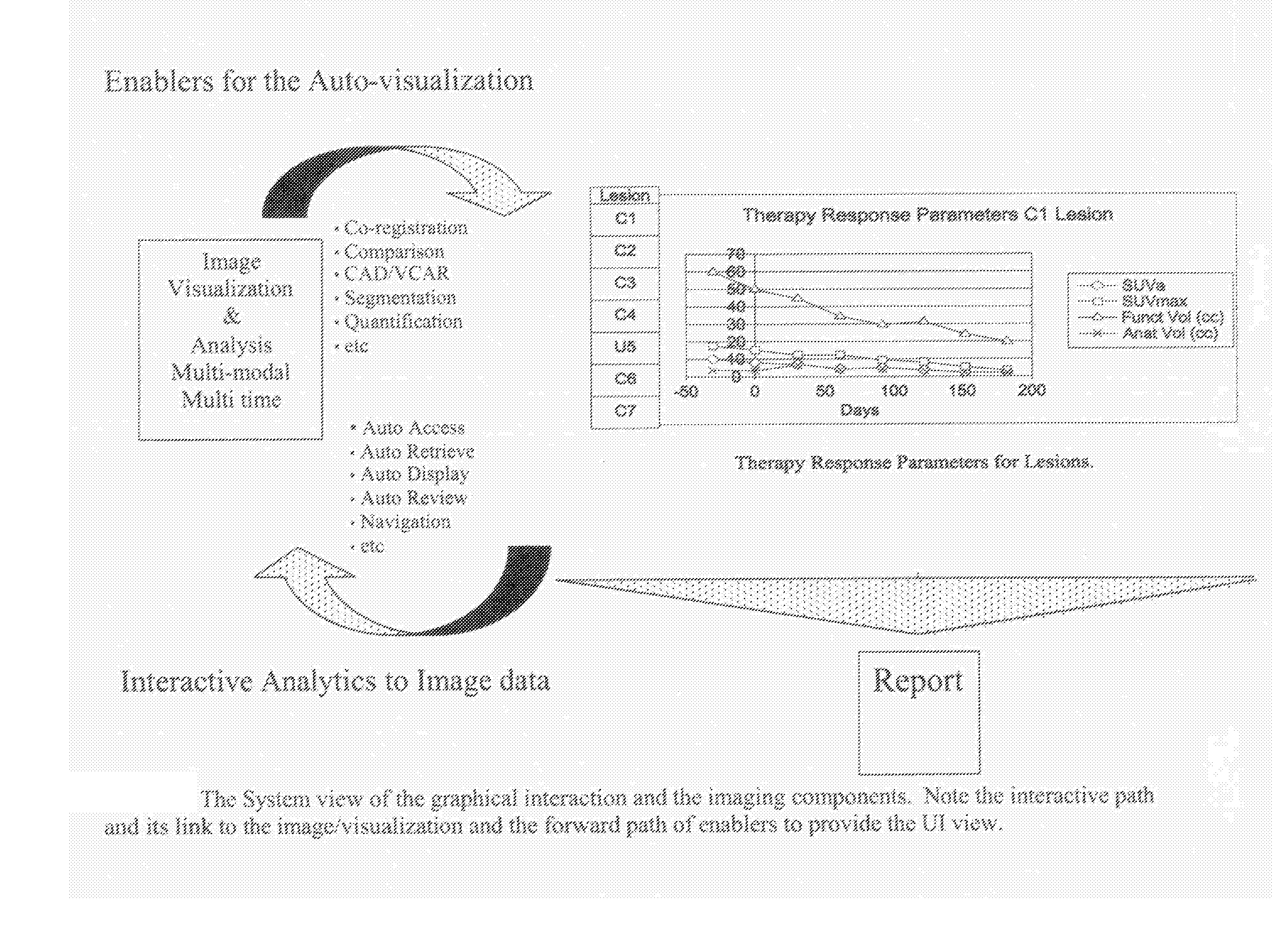
[0015]In yet another aspect, a method includes providing a direct interaction with therapy response parameters to facilitate a user's efficient analyzing of multi-modality and multi-time points exams.

Problems solved by technology

This is resulting in enormous numbers of CT slices to be inspected in soft-tissue, lung, mediastinal, abdominal, and bone CT windows.
This is a tedious and time-consuming task.

Method used


Image

  • Methods and Apparatus for Volume Computer Assisted Reading Management and Review
  • Methods and Apparatus for Volume Computer Assisted Reading Management and Review
  • Methods and Apparatus for Volume Computer Assisted Reading Management and Review

Examples


examples

[0147]In a PET / CT exam, the two exams are registered. For a given organ, both anatomical information (from the CT exam) and functional information (from the PET exam) are displayed together. This includes showing a fused image and reporting. See bottom right of FIG. 5 for a fused image.[0148]Two chest x-ray exams taken at different times are registered. For a given nodule, an image may display the differences in nodule size.[0149]In neurology, two MR exams are taken at different times on a patient with Alzheimer's. A difference image depicts disease progression over time.

[0150]Analysis may be in the form of measurements (depicted graphically or in text). Analysis displayed may be acquired from a single exam, multiple exams or the combination or exams.

[0151]Therapy Parameter Display

[0152]Therapy Parameter Display is the novel idea that will allow clinicians to interact with quantitative patient information, providing the ability to view the data analysis in graphical layouts, interac...


PUM


Abstract

A method includes providing an auto visualization display based on at least one quantitative analysis of at least one object of interest's progress over time regarding therapy response parameters over time.

Description

CROSS REFERENCE TO RELATED APPLICATIONS[0001]This application claims the benefit of U.S. provisional application Ser. No. 60 / 810,199 filed Jun. 1, 2006.BACKGROUND OF THE INVENTION[0002]This invention relates generally to diagnostic imaging methods and apparatus, and more particularly to methods and apparatus that provide volume computer assisted reading management and review (VCAR) tools for the purpose of displaying and managing therapy parameters and / or tumor responses to treatment over time. This disclosure is useful for all medical imaging modalities such as, for example, CT, MR, CT / PET, SPECT, X-Ray, and / or Ultrasound.[0003]A tumor is a cluster of cancer cells that are descendants of a single cell that underwent a malignant transformation. The increased growth rate of cancer cells results in an equally increased metabolic activity of these clusters.[0004]Over time the tumor volumes will increase in such a way that anatomical / morphological changes in or around the affected organ...

Claims


Application Information

Patent Timeline
Patent Type & Authority Applications(United States)
IPC IPC(8): A61B5/05
CPCG06T2207/30024G06T7/0012
Inventor GONZALEZ, MARCELA ALEJANDRABECKETT, BOB LOUISSIROHEY, SAAD AHMEDCONRY, ANNE MARIEAVINASH, GOPAL B.VAN NUFFEL, ANDRE JOHN PIERRE
Owner GENERAL ELECTRIC CO
Learn More