Assay for simultaneous genomic and proteomic analysis

a technology of simultaneous genomic and proteomic analysis, applied in the direction of microbiological testing/measurement, biochemistry apparatus and processes, etc., can solve the problems of small size, heterogeneous cellular content, difficult to obtain clinical samples especially in immuno-oncology areas, etc., and achieve high throughput screening and efficiently capture and analyze cells

Inactive Publication Date: 2018-07-26
MERCK SHARP & DOHME CORP
View PDF0 Cites 65 Cited by
  • Summary
  • Abstract
  • Description
  • Claims
  • Application Information

AI Technical Summary

Benefits of technology

[0004]The invention is a biochemical assay for simultaneous genomic and proteomic analysis. In one embodiment, it is used for single-cell resolution using sequencing as a readout. This method enables quantification of an unlimited number of proteins and RNA species in thousands of single-cells. In addition, this assay can also be applied to small subpopulations of cells or cell lysates for high throughput screening. The assay is designed to efficiently capture and analyze cells from small clinical biopsies where cell number is limited.

Problems solved by technology

Currently, clinical samples especially in the immuno-oncology area are difficult to obtain, heterogeneous in cellular content, and typically very small in size.
Unfortunately, clinical samples are typically limiting and there is insufficient material to perform both proteomic and genomic profiling.
This hybridization based method has limitations on multiplexing capabilities (1000's of cells).
However, the increase was a modest two-fold (˜37 markers) and since the cells are ionized in the process it does not allow for any further downstream analysis to be conducted such as RNA-sequencing.

Method used


Image

  • Assay for simultaneous genomic and proteomic analysis
  • Assay for simultaneous genomic and proteomic analysis
  • Assay for simultaneous genomic and proteomic analysis

Examples


example 1

of Protein Detection Probes

[0022]Antibodies are conjugated to nucleotides via commercially available kits such as Thunder-link by Innova Biosciences (Lundberg et al., 2011a) and the antibody-oligonucleotide all-in-one kit by Solulink. Solulink's technology is based on the use of two complementary heterobifunctional linkers. It uses a two-step process to prepare an antibody-oligonucleotide. In the first step, an amine-modified 20 to 60 mer oligonucleotide is modified using an excess of Sulfo-S-4FB linker or by solid phase oligonucleotide synthesis using 4FB-phosphoramidite (Kasai et al., 2012). This reactive NHS-ester incorporates a 4FB (aromatic aldehyde functional group, formylbenzamide) at the desired terminus (5′ or 3′ end) of the oligonucleotide. In the second step, polyclonal or monoclonal antibodies are modified with S-HyNic linker. This NHS-ester reacts with lysine residues on the antibody incorporating HyNic functional groups (hydrazino-nicotinamide). The HyNic-modified anti...

example 2

Protein Molecules with Target Probes

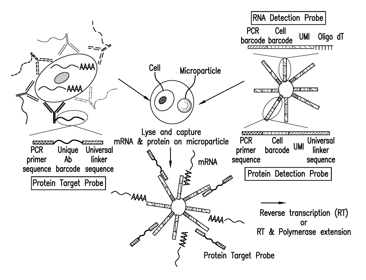
[0028]Protein molecules on the cell surface of living cells are labeled with antibodies conjugated to oligonucleotides or aptamers. To label intracellular protein molecules the cells are fixed and permeabilized. Then antibodies conjugated to oligonucleotides or aptamers are added. Cells are centrifuged and supernatant with excess unbound antibodies or aptamers is removed. Washes are conducted to further remove unbound antibodies and aptamers. Cells are isolated and protein detection probes are added that will bind to the protein target probes.

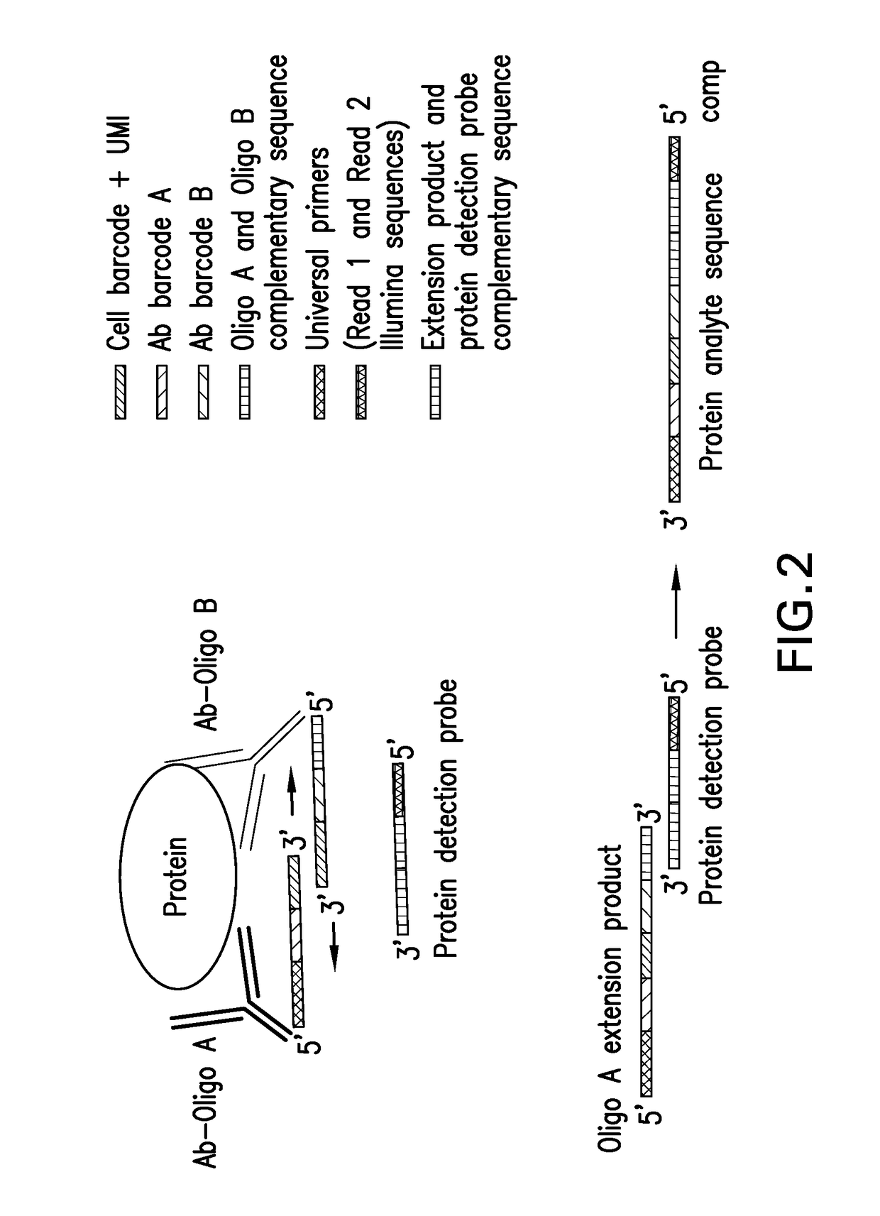
[0029]Alternatively, cells are lysed and extracellular and intracellular protein molecules are in solution. For each protein molecule of interest two antibodies, each conjugated with its own oligonucleotide with a complementary sequence to each other is added. A signal is produced only when the two antibodies bind to the same protein molecule and bring the two oligonucleotides in proximity, and form a DNA tem...

example 3

RNA Molecules with Detection Probes

[0030]Detection probes containing a poly dT sequence capture the polyA tail of the mRNA. The mRNA is reverse transcribed to cDNA and a template switching oligonucleotide (TSO) is used to introduce a PCR handle downstream of the synthesized cDNA (Zhu et al., 2001). Exonuclease 1 (NEB) is added to remove excess unbound detection probes. The cDNA is PCR amplified and then is quantified. The cDNA is fragmented and amplified for sequencing with the Nextera XT DNA sample prep kit (Illumina) using custom primers that enabled the specific amplification of only the 3′ ends (Macosko et al., 2015).

[0031]Alternatively, library preparation is based on the CEL-Seq protocol with minor modifications (Jaitin et al., 2014). The workflow of DNA library preparation is summarized as follows: reverse transcription (RT)→Exonuclease I→SPRI purification (SPRIP)→Second Strand Synthesis (SSS)→SPRIP→T7 in vitro transcription linear amplification→SPRIP→RNA Fragmentation→SPRIP→...


PUM

PropertyMeasurementUnit
specific diameteraaaaaaaaaa
lengthaaaaaaaaaa
sizeaaaaaaaaaa

Abstract

The present invention is directed to a biochemical assay for simultaneous genomic and proteomic analysis.

Description

BACKGROUND OF THE INVENTION[0001]Currently, clinical samples especially in the immuno-oncology area are difficult to obtain, heterogeneous in cellular content, and typically very small in size. Often times, for a given sample genomic profiling, proteomic analysis, immune profiling, biomarker monitoring etc. is needed. Unfortunately, clinical samples are typically limiting and there is insufficient material to perform both proteomic and genomic profiling. There are many post translational modifications that occur to proteins such as phosphorylation, ubiquitination, glycosalation, etc. that play an important role in regulating different cellular signaling pathways. There is a need for assays that enables quantification of an unlimited number of proteins and RNA species at the single-cell level.[0002]Proof of concept of labeling cellular proteins with antibodies attached to DNA has been previously demonstrated using the NanoString platform as a readout (Ullal & Peterson, Sci. Transl. M...

Claims


Application Information

Patent Timeline
Patent Type & Authority Applications(United States)
IPC IPC(8): C12Q1/6811C12Q1/6806C12Q1/6837C12Q1/6851C12Q1/686C12Q1/6876
CPCC12Q1/6811C12Q1/6806C12Q1/6837C12Q1/6851C12Q1/686C12Q1/6876C12Q2600/158C12Q2600/166C12Q2600/16C12Q2521/107C12Q2563/149C12Q2563/179C12Q2565/537
Inventor PETERSON, VANESSA MARIEKLAPPENBACH, JOEL ALBERT
Owner MERCK SHARP & DOHME CORP
Why Eureka
Try Eureka