CMOS image sensors adapted for dental applications

a technology of image sensors and dental examinations, applied in the field of dental examinations, can solve the problems of increasing the cost of dental examination, time, cost, equipment needed to process the film, and patient exposure to a significant dose of x-rays

Active Publication Date: 2008-01-31
BAE SYST IMAGING SOLUTIONS
View PDF19 Cites 12 Cited by
  • Summary
  • Abstract
  • Description
  • Claims
  • Application Information

AI Technical Summary

Benefits of technology

[0005]The present invention includes an image sensor having a two-dimensional array of CMOS pixel sensors, a row decoder and a column decoder. The two-dimensional array of CMOS pixel sensors array are organized as a plurality of rows and columns. The pixel sensors in each column are connected to a corresponding bit bus by a row select switch that connects the pixel sensor in the row to that bit bus in response to a row select signal on a corresponding row select line. The row decoder generates the row select signal on a selected one of the row select lines in response to a row address that is coupled thereto. The column decoder connects a selected one of the bit buses to an interface connecting the image sensor to a circuit that is external to the image sensor. One of the column decoder or the row decoder is located between two of the rows or two of the columns, respectively. X-rays are converted to light that is detected by the image sensor by a layer of scintillation material that overlies the two-dimensional array. In one aspect of the invention, the two-dimensional array includes a rectangular array having a chamfered corner such that the rows or columns that extend into the chamfered corner have lengths that are less than the rows or columns, respectively, that do not extend into the corners.

Problems solved by technology

While this method has been in use for many years, it has its disadvantages.
First, the patient is exposed to a significant dose of x-rays.
Second, the time, cost, and equipment needed to process the film increases the cost of the dental examination.
Third, the chemicals utilized in processing the film pose a disposal problem.
These problems have led to several attempts to replace the film component of the traditional x-ray examination with a solid-state sensor that is placed in the patient's mouth to record the x-ray image.
Unfortunately, conventional solid-state imaging sensors have a shape that is predominately rectangular.
However, sensors based on CCD arrays are less than ideal for intra-oral x-ray sensors.
In addition, CCD arrays require relatively high voltages to operate and are more expensive to fabricate than CMOS sensors.
Since cost is an important factor in achieving acceptance of a new imaging system by the dental community, these additional costs can be a serious barrier to the introduction of solid-state imaging systems.

Method used


Image

  • CMOS image sensors adapted for dental applications
  • CMOS image sensors adapted for dental applications
  • CMOS image sensors adapted for dental applications

Examples


Embodiment Construction

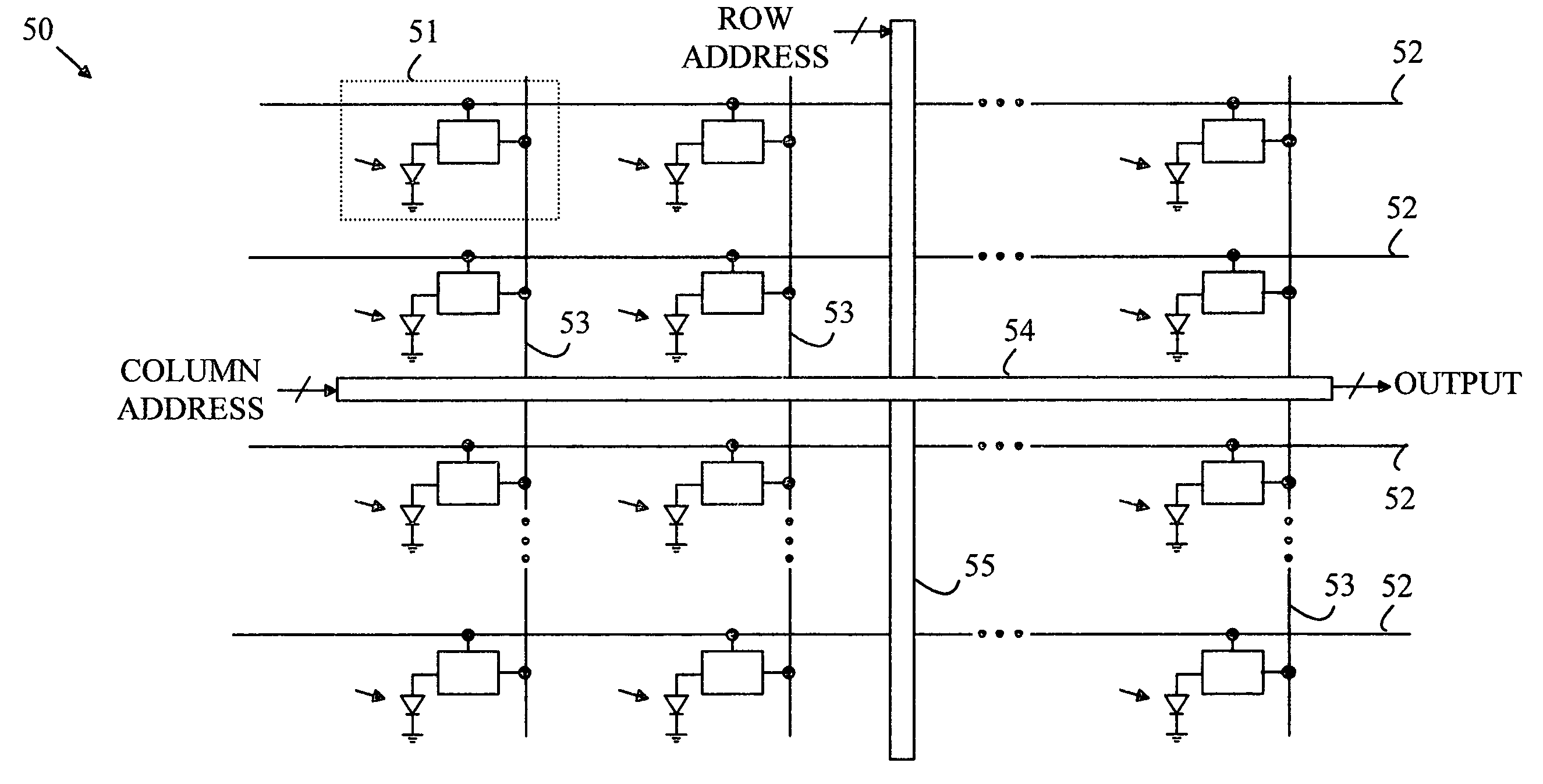
[0017]The manner in which the present invention provides its advantages can be more easily understood with reference to FIG. 1, which is a block diagram of a prior art conventional CMOS image sensor. CMOS image sensor 20 includes a two-dimensional pixel array 21, which is the active area for image measurement, and some peripheral circuits that do not contribute to image sensing. These,circuits generally include a logic controller 24, and row and column decoders 22 and 23. The individual pixels are read out over bit lines that terminate in a row decoder / sense amplifier. Sensor die 25 is normally rectangular, and the pixel array normally does not extend to all four edges of the die.

[0018]Light from the image generates a charge signal inside each pixel. After a fixed integration time, the pixel charge signals are read out from the array and are eventually digitized to form a digital image. The readout of the pixel array is facilitated by the row and column decoders, and operates much l...


PUM


Abstract

An image sensor having a two-dimensional array of CMOS pixel sensors, a row decoder and a column decoder is disclosed. The two-dimensional array of CMOS pixel sensors is organized as a plurality of rows and columns that are addressed with the aid of row and column decoders. At least one of the column decoder or the row decoder is located between two of the rows or two of the columns, respectively. X-rays are converted to light that is detected by the image sensor by a layer of scintillation material that overlies the two-dimensional array. The internally located decoder or decoders facilitate sensors in which the two-dimensional array includes a rectangular array having a chamfered corner such that rows or columns that extend into the chamfered corner have lengths that are less than rows or columns, respectively, that do not extend into the corners.

Description

BACKGROUND OF THE INVENTION[0001]Dental x-rays are typically taken with a film that is placed in the patient's mouth. The film is exposed through the teeth by an x-ray source that resides outside the patient's head. While this method has been in use for many years, it has its disadvantages. First, the patient is exposed to a significant dose of x-rays. This dose is accumulative over the patient's lifetime. Second, the time, cost, and equipment needed to process the film increases the cost of the dental examination. Third, the chemicals utilized in processing the film pose a disposal problem.[0002]These problems have led to several attempts to replace the film component of the traditional x-ray examination with a solid-state sensor that is placed in the patient's mouth to record the x-ray image. In such systems, a layer of scintillation material is used to convert the x-rays to visible light. The visible light is then imaged onto a solid-state imaging array. Since solid-state x-ray s...

Claims


Application Information

Patent Timeline
Patent Type & Authority Applications(United States)
IPC IPC(8): H04N5/335A61B6/14H04N5/32H04N5/374H04N5/376
CPCH04N5/32H04N5/335A61B6/145G01T1/2018H04N5/374H04N25/00H04N25/76G01T1/20184G01T1/2006
Inventor LIU, XINQIAOFOWLER, BOYD
Owner BAE SYST IMAGING SOLUTIONS
Why Eureka
Try Eureka