Method for Creating 3D Coordinate Systems in Image Space for Device and Patient Table Location and Verification

a technology of image space and coordinate system, applied in the direction of patient positioning for diagnostics, therapy, application, etc., can solve the problem of missing the tumor entirely, and achieve the effect of accurately targeting the lesion and accurately positioning the patien

Inactive Publication Date: 2008-02-07
QFIX SYSTEM LLC
View PDF18 Cites 93 Cited by
  • Summary
  • Abstract
  • Description
  • Claims
  • Application Information

AI Technical Summary

Benefits of technology

[0008] The instant invention also provides a method of accurately positioning a patient on a couch top or device taking into account deformation of the couch top or device due to the weight of the patient.
[0009] The instant invention also provides a method for accurately targeting a lesion during radiation treatment through image guidance comprising determining location of one or more image markers in real time using at least one selected from the group consisting of lasers, visual, infrared, MRI, RF and radiation; and modifying at least one of the patient position or radiation treatment beam path to adaptively compensate for a change in position.

Problems solved by technology

If a highly tailored beam is off target by a few millimeters, it may miss the tumor entirely.

Method used


Image

  • Method for Creating 3D Coordinate Systems in Image Space for Device and Patient Table Location and Verification
  • Method for Creating 3D Coordinate Systems in Image Space for Device and Patient Table Location and Verification
  • Method for Creating 3D Coordinate Systems in Image Space for Device and Patient Table Location and Verification

Examples


Embodiment Construction

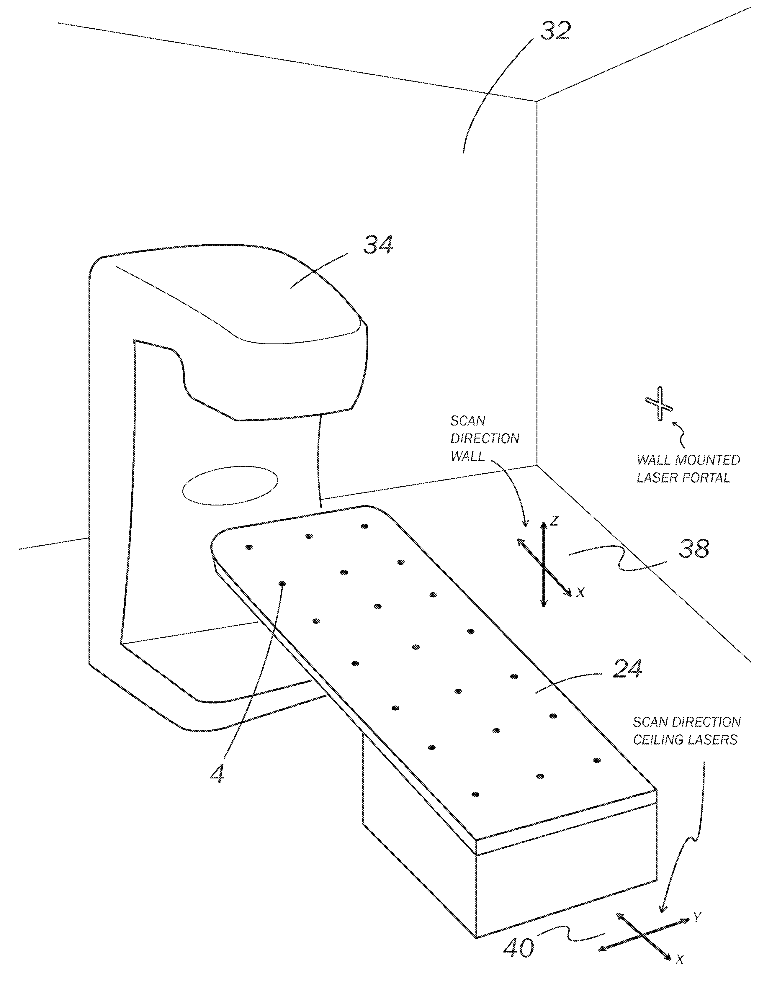
[0022] Both in simulation and treatment, it is desirable to know that the couch top, devices, and patient are in the proper position. This starts at the point of simulation in which the patient is scanned using conventional x-ray, CT, MRI, radio-frequency, PET, SPECT or other modalities to determine the location of the cancerous lesion. Because radiation therapy is often delivered in multiple fractions, it is important to be able to confirm the location of the patient accurately and repeatably.

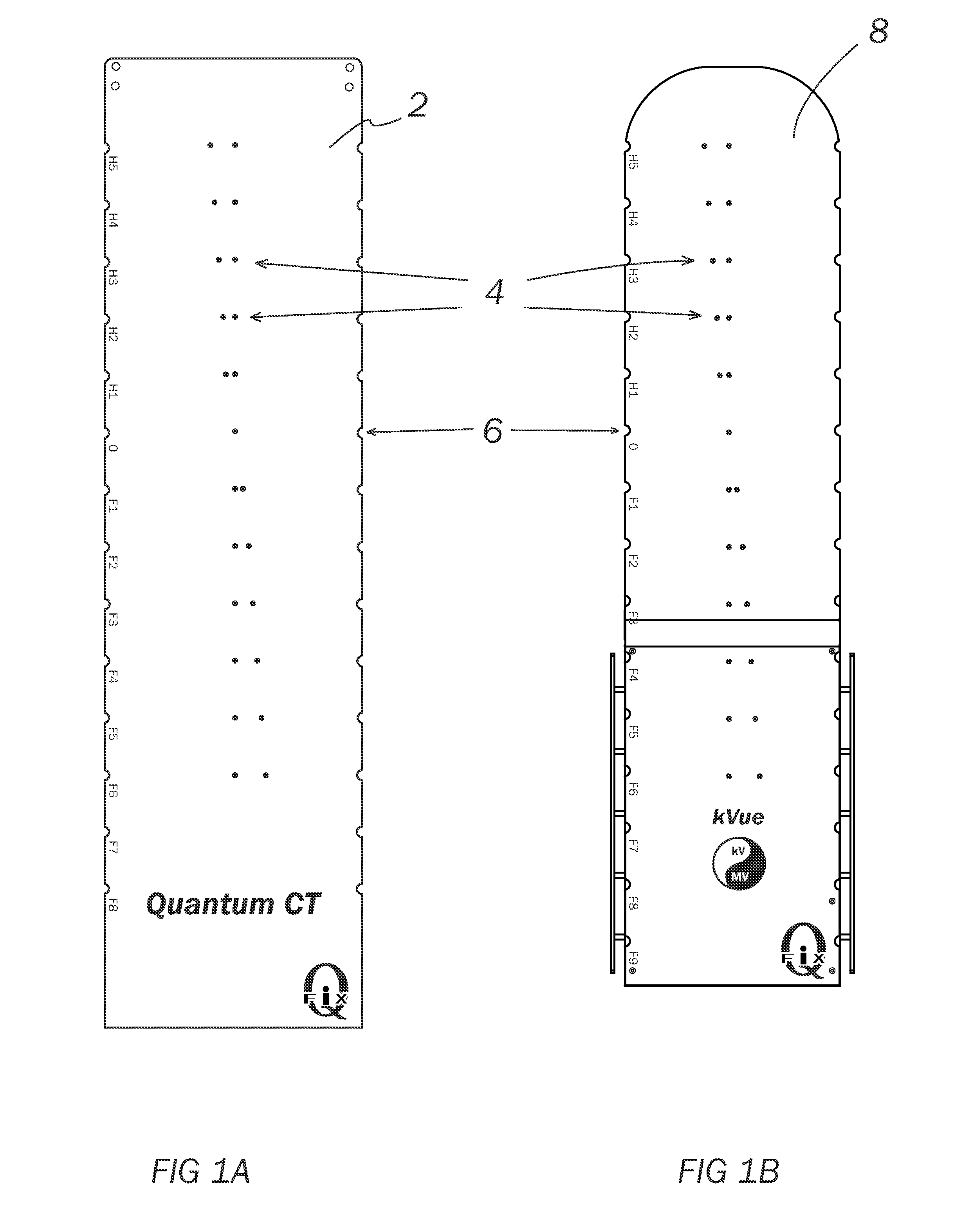
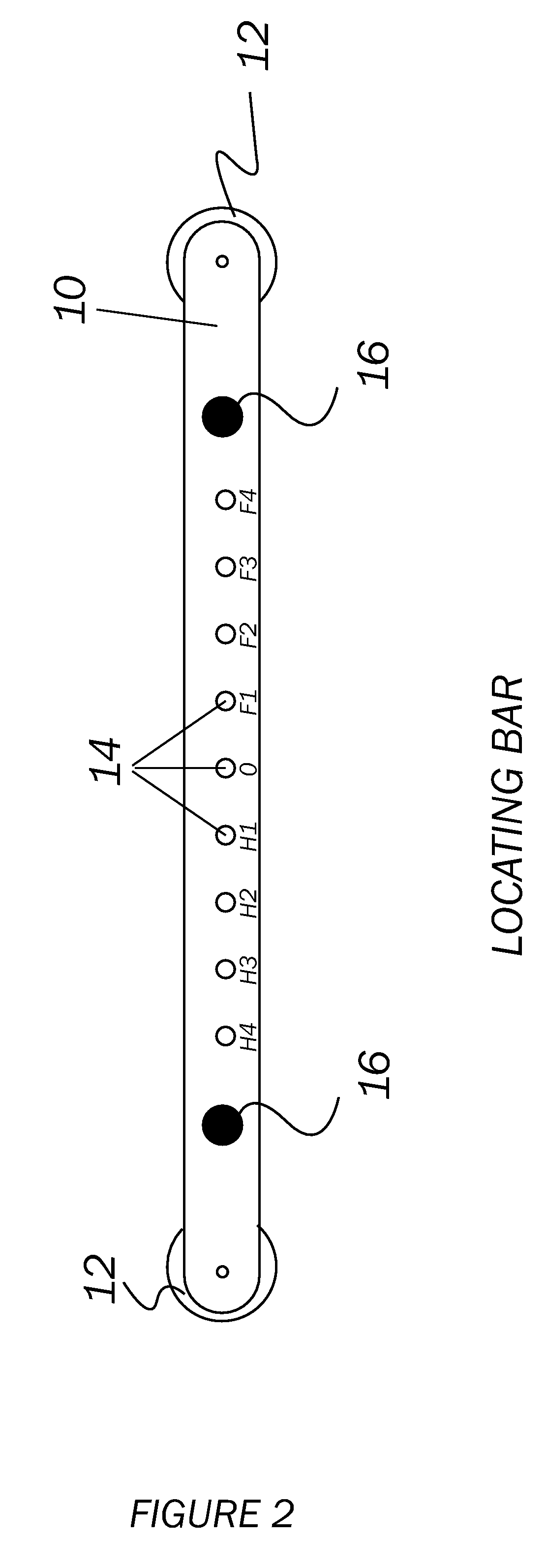
[0023] The incorporation of diagnostic imaging tools directly on the radiation therapy treatment machine (be it a LINAC, proton therapy or other variety) means that markers can now be used to identify the location of the patient positioning devices and table top. Continuous markers in the form of a pair of diverging lines have been used to provide an axial location on CT scanners for years. However, they do not allow the user to accurately locate specific positions. Physical patient positioni...


PUM


Abstract

The present invention provides a patient couch top or device for quickly and accurately positioning a patient during simulation and treatment by placing a series of small fiducial markers in discrete locations on the couch top or device. With use of the fiducial markers, the present invention allows for the correction for misalignment and deformation of patient positioning equipment which occurs due in part to a patient's size and weight. The present invention also provides a method for positioning a patient and correcting for deformation of the couch top or device.

Description

[0001] This application claims priority to and benefit of U.S. Provisional Application No. 60 / 795,836 filed 27 Apr. 2006, entitled Radiation Therapy Patient Couch Top Compatible with Diagnostic Imaging.BACKGROUND OF THE INVENTION [0002] State of the art cancer radiation therapy is increasingly based on the pin point application of high energy radiation which is highly tailored to the shape and position of the cancerous tumor. Modern techniques such as IMRT use a pencil sized beam whose cross-section is shaped to match the tumor. This allows the physician to spare the surrounding healthy tissue while increasing the treatment dose to the cancerous target. As the size of the treatment beam decreases, the accurate location of the beam becomes much more critical. If a highly tailored beam is off target by a few millimeters, it may miss the tumor entirely. [0003] Because of these new techniques, it becomes increasingly desirable to know the position and shape of the tumor accurately with ...

Claims


Application Information

Patent Timeline
Patent Type & Authority Applications(United States)
IPC IPC(8): A61N5/10A61B6/04A61B6/08
CPCA61B6/04A61B6/0492A61B6/08A61N2005/1061A61N5/1049A61B5/05
Inventor COPPENS, DANIEL D.
Owner QFIX SYSTEM LLC
Why Eureka
Try Eureka