Rapid and sensitive assay for the detection and quantification of coregulators of nucleic acid binding factors

a nucleic acid binding factor and coregulator technology, applied in the field of biosensors, can solve the problems of laborious and time-consuming methods, and the general adaptability of methods to high-throughput assay formats, and achieve the effect of facilitating or stabilizing the spectroscopic interaction and measurable changes in fluorescen

Inactive Publication Date: 2008-02-21
SAINT LOUIS UNIVERSITY
View PDF19 Cites 10 Cited by
  • Summary
  • Abstract
  • Description
  • Claims
  • Application Information

AI Technical Summary

Benefits of technology

[0023] In another embodiment, a single polynucleotide is labeled in two positions, with a fluorescent donor at the first position and with a fluorescent acceptor at the second position, wherein the fluorescent labels are at such a distance from one another so as not to interact spectroscopically in the absence of a bridging nucleic acid binding factor. In one aspect of this embodiment a portion (component) of a nucleic acid element is located near the first position and another portion (component) of the same nucleic acid element is located near the second position. Upon the binding of a nucleic acid binding factor to both portions of said nucleic acid element, the first position is brought into proximity to the second position, thereby facilitating or stabilizing the spectroscopic interaction between fluorescent donor and fluorescent acceptor. In another aspect of this embodiment, a first complete nucleic acid element is located at or near the first position and a second complete nucleic acid element is located at or near the second position. Upon the binding of a nucleic acid binding factor or complex assembly of nucleic acid binding factors (as in an enhanceosome, for example) to the first and second element, the first position is brought into proximity to the second position, thereby facilitating or stabilizing the spectroscopic interaction between fluorescent donor and fluorescent acceptor, resulting in a measurable change in fluorescence due to fluorescent energy transfer or quenching.

Problems solved by technology

These methods are laborious and time-consuming procedures, which typically involve the use of dangerous and expensive radioisotopes.
Furthermore, these methods are not generally adaptable to high-throughput assay formats.

Method used


Image

  • Rapid and sensitive assay for the detection and quantification of coregulators of nucleic acid binding factors
  • Rapid and sensitive assay for the detection and quantification of coregulators of nucleic acid binding factors
  • Rapid and sensitive assay for the detection and quantification of coregulators of nucleic acid binding factors

Examples


example 1

Detection of cAMP Receptor Protein (CAP), a Sequence-Specific Nucleic Acid Binding Protein from E. coli

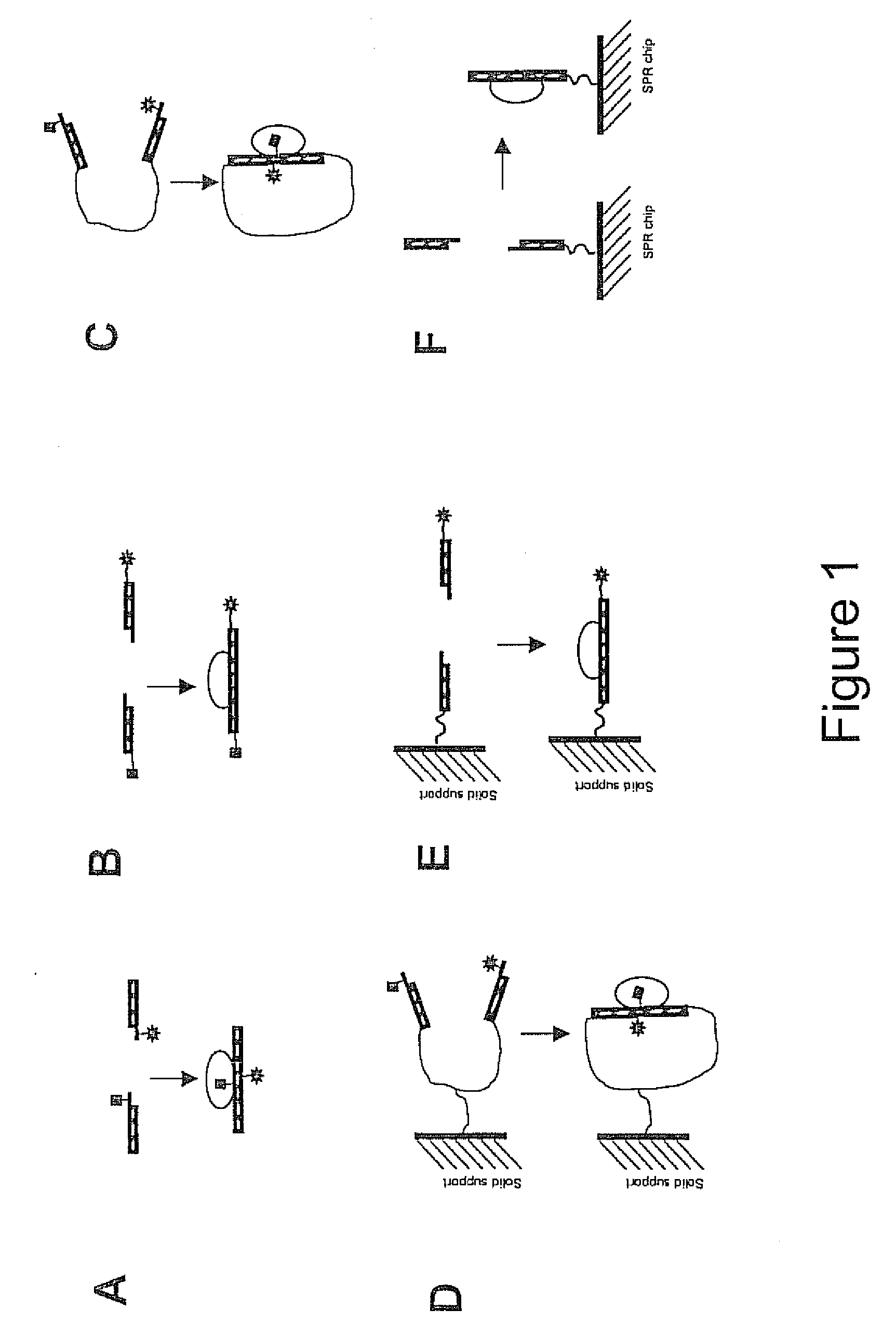
[0123] CAP is a bacterial transcription activator which binds DNA at a Kd=˜0.1 nM in a sequence specific manner (Busby, S., and Ebright, R. H. J. Mol. Biol. 293, 199-213, 1999). A 38 bp DNA sequence corresponding to a consensus CAP site (Ebright, R. H., Ebright, Y. W. & Gunasakera, A. Nucleic Acids Res. 17, 10295-10305, 1989) was used as a basis for designing oligonucleotides necessary for preparing the CAP assay reagents according to the scheme illustrated in FIG. 1A. FIG. 3 illustrates the details of the design used. The following four oligonucleotides were synthesized using standard phosphoramidate automated oligonucleotide synthesis (F=dT-fluorescein; D=dT-dabcyl):

5′-AACGCAATAAATGTGA(CAP1; SEQ ID NO:1)5′-AGFAGATCACATTTTAGGCACC 3′(CAP2; SEQ ID NO:2)5′-GGTGCCTAAAATGTGA(CAP3; SEQ ID NO:3)5′-TCDACTTCACATTTATTGCGTT(CAP4; SEQ ID NO:4)

[0124] The fluorescence donor (fluorescein) and...

example 2

Demonstration of the Use of Fluorochromes with Different Emission Spectra and Different Modes of Fluorescence Signal Detection

[0131] The following oligonucleotides, which have identical sequences to CAP2 and CAP4, respectively, were synthesized using standard phosphoramidate automated oligonucleotide synthesis, wherein X represents amino-dT:

5′-AGXAGATCACATTTTAGGCACC-3′(CAPS; SEQ ID NO:7)5′-TCXACTTCACATTTATTGCGTT-3′(GAPS; SEQ ID NO:8)

[0132] Amino-Modifier C2 dT (Glen Research, Sterling, Va.) was incorporated into positions that are equivalent to the positions at which fluorescein-dt and dabcyl-dT has been used previously in CAP2 and CAP4 oligonucleotides, respectively. Amino-Modifier C2 dT contains a reactive aliphatic amino group that can be used to covalently attach any aminoreactive fluorescence probe. CAP5 (SEQ ID NO:7) and CAP6 (SEQ ID NO:8) oligonucleotides were modified with 7-diethylaminocoumarin-3-carboxylic acid, succinimidyl ester. This fluorochrome is excited at 433 nm...

example 3

The Detection of Nucleic Acid Binding Protein Coregulators, for Example cAMP

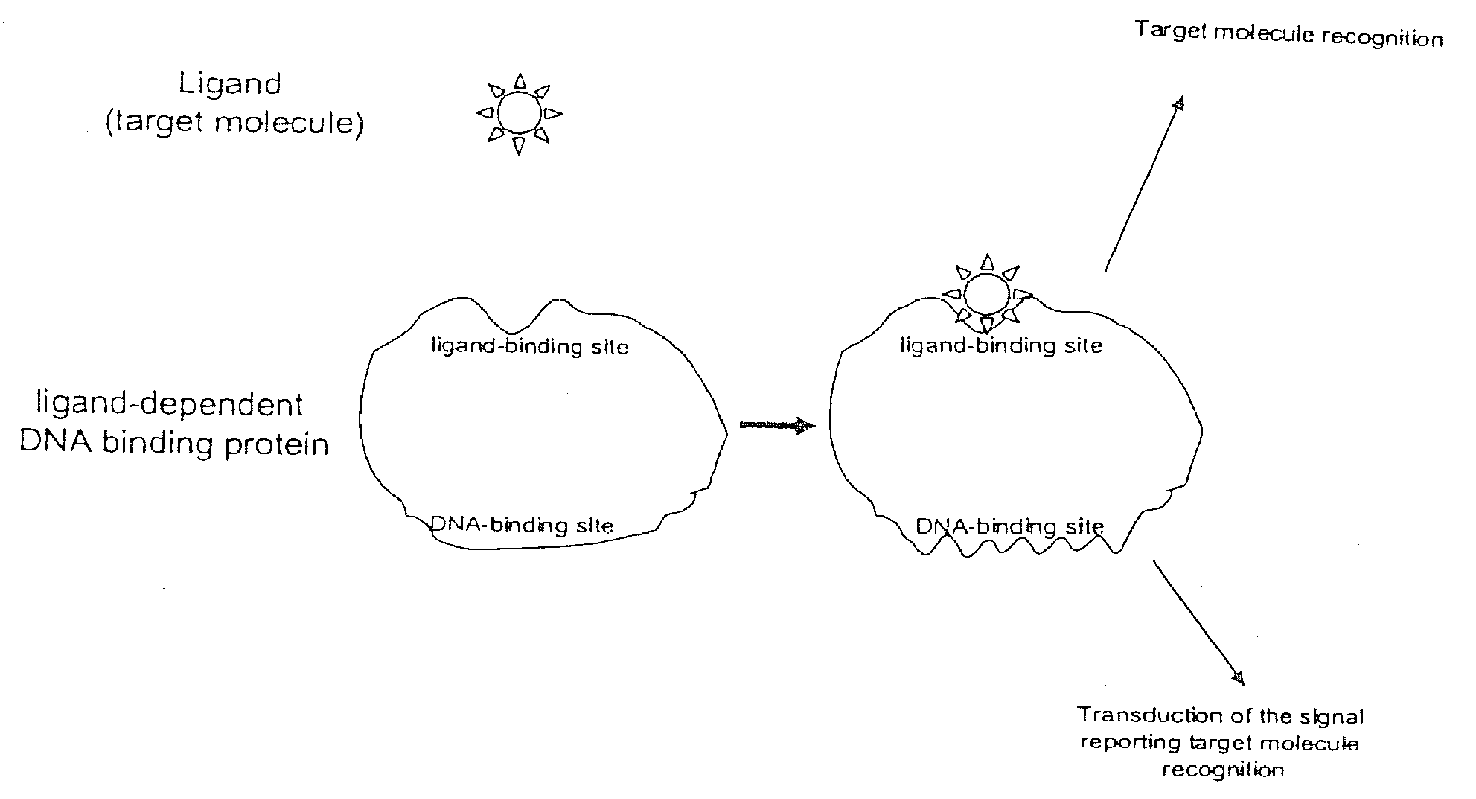
[0135] The activity of many nucleic acid binding proteins is regulated by small molecules, other proteins or cellular events (e.g., phosphorylation). Hence, the present invention may be used to identify or detect these regulatory molecules or regulatory cellular events.

[0136] CAP protein binds both cAMP and cGMP with micromolar affinity (Takahashi, M., Blazy, B., and Baudras, A. Biochemistry 19, 5124-30). Thus, CAP protein, by itself, is a poor reagent for specifically detecting cAMP. However, the high affinity of CAP for its cognate nucleic acid binding sequence is selectively dependent on cAMP, but not cGMP. Since only cAMP is thermodynamically linked to sequence-specific nucleic acid binding, in the presence of DNA containing a CAP binding site, the affinity of CAP for cAMP is increased about 1000-fold, whereas the affinity for cGMP remains unchanged. Thus, in the presence of DNA containing a CAP bindin...


PUM

PropertyMeasurementUnit
excitation wavelengthaaaaaaaaaa

Abstract

Biosensors and methods to determine the activity of any and all nucleic acid binding factors, proteins, cellular events, nucleic acid binding protein coregulators, or fragments thereof, based upon the stabilization of the interaction of two nucleic acid components, which together comprise a complete nucleic acid binding element, by the binding of a nucleic acid binding factor are provided. Preferably, a fluorescence donor is attached to a nucleic acid comprising one portion or component of a complete nucleic acid binding element and a fluorescence acceptor is attached to a nucleic acid comprising the other portion or component of the same complete binding element. Alternatively, a solid substrate is attached to a nucleic acid comprising one portion of a binding element and a detectable label is attached to a nucleic acid comprising the other portion of the same binding element. Binding of a nucleic acid binding factor to the nucleic acid components affects a change in luminescence or the association of the detectable label with the solid substrate. These biosensors and methods may also be used to detect mediating nucleic acid binding factor coregulators, post-translational modifications and cellular events, to diagnose diseases and / or screen for drugs or other ligands that mediate the activity of nucleic acid binding factors.

Description

CROSS REFERENCE TO RELATED APPLICATIONS [0001] This application is a divisional of copending U.S. patent application Ser. No. 10 / 062,064, filed Jan. 31, 2002, the disclosure of which is hereby incorporated by reference, which is a continuation-in-part of U.S. patent application Ser. No. 09 / 928,385, now U.S. Pat. No. 6,544,746, filed on Aug. 13, 2001, the disclosure of which is also hereby incorporated by reference.GOVERNMENTAL SUPPORT [0002] This work was supported by the U.S. Department of Health and Human Services / National Institutes of Health grant number GM50514. The U.S. Government has certain rights in this invention.SEQUENCE LISTING [0003] A paper copy of the sequence listing and a computer readable form of the same sequence listing are appended below and herein incorporated by reference. The information recorded in computer readable form is identical to the written sequence listing, according to 37 C.F.R. 1.821 (f). BACKGROUND OF THE INVENTION [0004] 1. Field of the Inventio...

Claims


Application Information

Patent Timeline
Patent Type & Authority Applications(United States)
IPC IPC(8): C12Q1/68
CPCG01N33/542G01N33/5308Y10S977/792
Inventor HEYDUK, TOMASZ
Owner SAINT LOUIS UNIVERSITY
Why Eureka
Try Eureka