Hood member for use with an endoscope

a technology for endoscopy and hood members, which is applied in the field of enhanced equipment, can solve the problems of destroying the histopathologic assessment of the lesion, emr procedures generally are not recommended, and the assessment of fragmented tissue may be more difficult than the assessment of unfragmented tissue,

Inactive Publication Date: 2008-03-06
COOK MEDICAL TECH LLC
View PDF19 Cites 103 Cited by
  • Summary
  • Abstract
  • Description
  • Claims
  • Application Information

AI Technical Summary

Benefits of technology

[0011]In accordance with one aspect, a portion of the lever portion is configured to be inserted beneath a section of mucosal tissue having a lesion during an ESD procedure. The lever p

Problems solved by technology

Further, assessment of fragmented tissue may be more difficult than assessment of unfragmented tissue.
One reported drawback associated with conventional EMR procedures is that the snaring method tends t

Method used


Image

  • Hood member for use with an endoscope
  • Hood member for use with an endoscope
  • Hood member for use with an endoscope

Examples


Embodiment Construction

[0029]In the present application, the term “proximal” refers to a direction that is generally towards a physician during a medical procedure, while the term “distal” refers to a direction that is generally towards a target site within a patent's anatomy during a medical procedure.

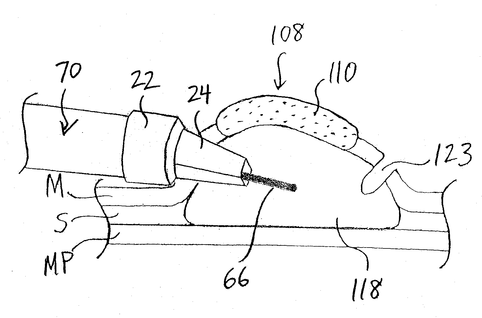
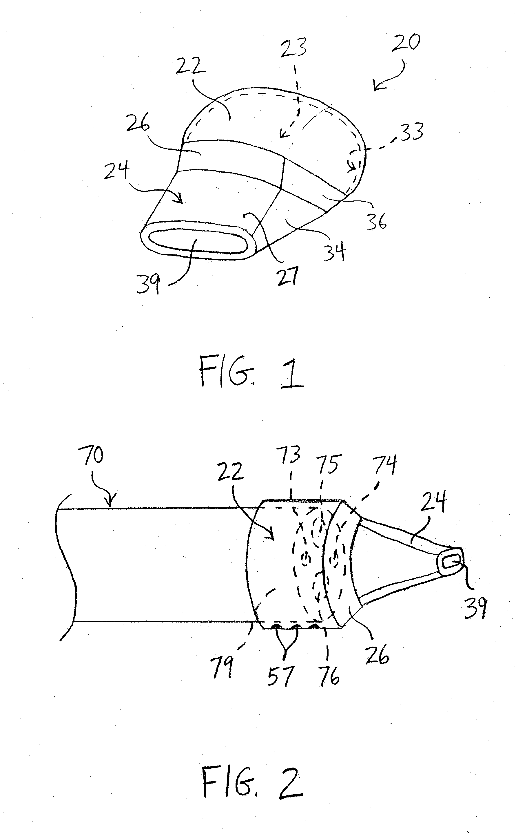
[0030]Referring now to FIG. 1, a first embodiment of a hood member of the present invention is shown. In FIG. 1, hood member 20 comprises hood portion 22 and lever portion 24. Hood portion 22 has interior surface 33 and hollow lumen 23 formed therein. Hood portion 22 may comprise a circular, oval or other configuration when viewed from the end (see, e.g., FIGS. 11A-11C below). As will be explained in greater detail below, hood portion 22 is adapted to be at least partially disposed over a distal region of a conventional endoscope, such as endoscope 70 of FIG. 2.

[0031]Referring still to FIG. 1, lever portion 24 preferably comprises a shape similar to a flat-head screwdriver. However, lever portion 24 may hav...


PUM


Abstract

The present invention provides apparatus and methods for performing endoscopic mucosal resection and endoscopic submucosal dissection of tissue. In a first embodiment, a hood member having a hood portion and a lever portion is provided. The hood portion is adapted to be disposed over a distal region of an endoscope. A portion of the lever portion is configured to be inserted beneath a section of mucosal tissue having a lesion, and the lever portion is configured to be rotated or otherwise maneuvered to lift the mucosal tissue in an upward direction, thereby facilitating removal of the tissue comprising the lesion. Optionally, a surgeon may advance a needle knife through the endoscope and lever portion to further incise submucosal tissue while the lever portion is disposed beneath the lesion. If desired, a flushing fluid may be provided to a target site during the procedure.

Description

PRIORITY CLAIM[0001]This invention claims the benefit of priority of U.S. Provisional Application Ser. No. 60 / 842,486, entitled “Hood Member For Use With an Endoscope,” filed Sep. 5, 2006, the disclosure of which is hereby incorporated by reference in its entirety.TECHNICAL FIELD[0002]The present invention relates generally to enhanced apparatus and methods for performing an endoscopic mucosal resection or submucosal dissection of tissue.BACKGROUND INFORMATION[0003]Diagnostic and therapeutic gastrointestinal endoscopy are commonly used to gain access to the digestive tract for the purpose of removing tissue. One technique for obtaining tissue for biopsies is an endoscopic mucosectomy procedure, also known as endoscopic mucosal resection (“EMR”). The EMR procedure may be a useful tool for providing a tissue specimen for surgical pathology.[0004]The EMR procedure also may be used for curative purposes to remove sessile benign tumors and intramucosal cancers, and in particular, EMR is ...

Claims


Application Information

Patent Timeline
IPC IPC(8): A61B1/005
CPCA61B1/00089A61B1/00101A61B2017/320044A61B17/320016A61B2017/00269A61B1/018
Inventor KARPIEL, JOHN A.
Owner COOK MEDICAL TECH LLC
Why Eureka
Try Eureka