
Dental x-ray film viewing device

a viewing device and x-ray technology, applied in the field of dental x-ray film viewing devices, can solve the problems of difficult diagnosis and patient case presentation of patients using x-ray films, the objects being radiographed, and the patients' teeth being also small,

Inactive Publication Date: 2008-03-27
LUCAS JAMES JOHN +2
View PDF7 Cites 1 Cited by
  • Summary
  • Abstract
  • Description
  • Claims
  • Application Information

AI Technical Summary

Benefits of technology

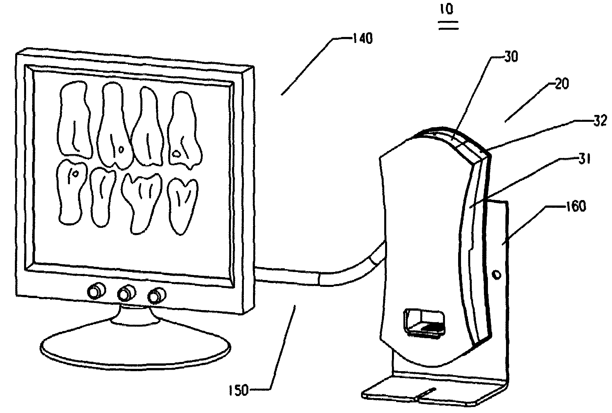
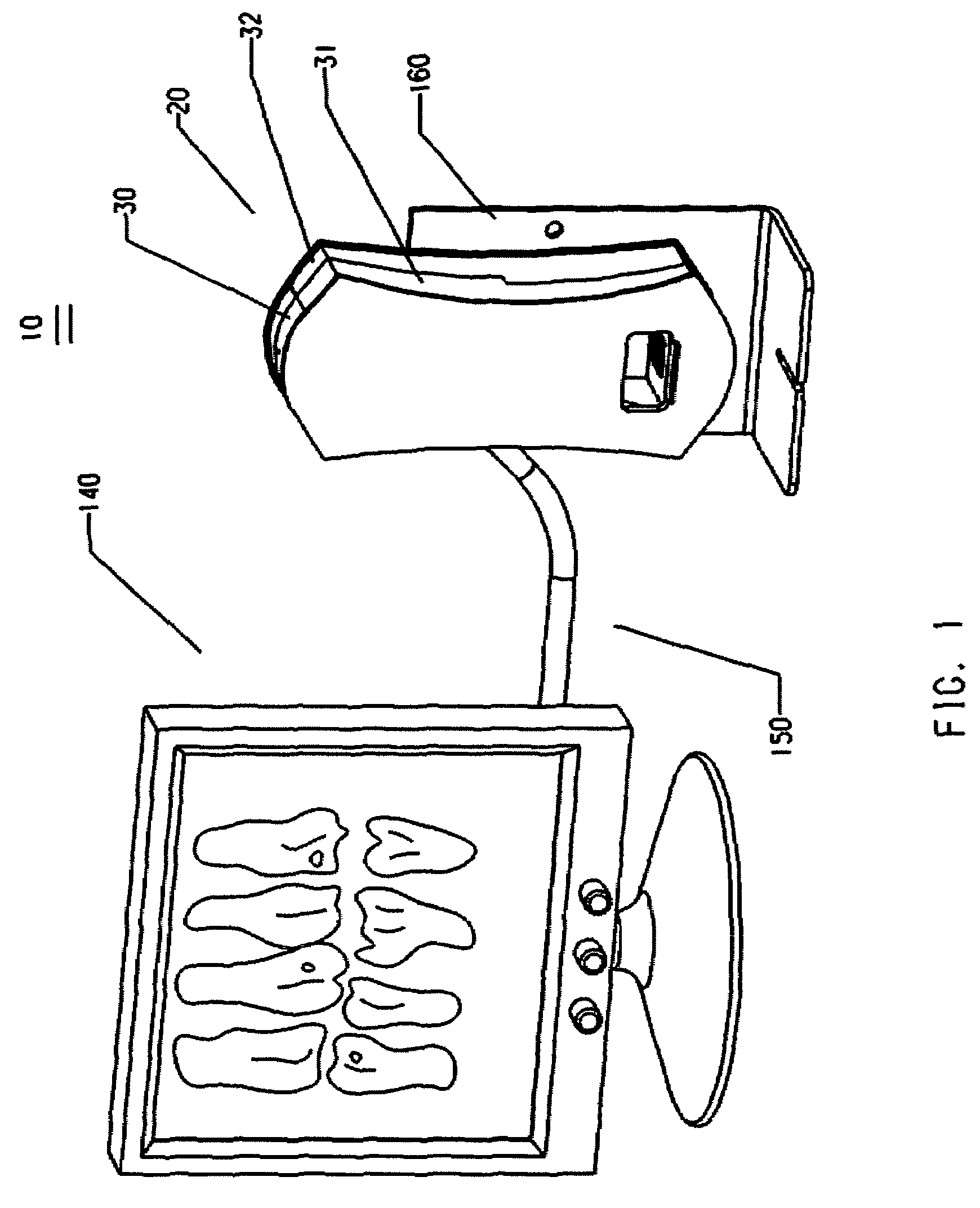
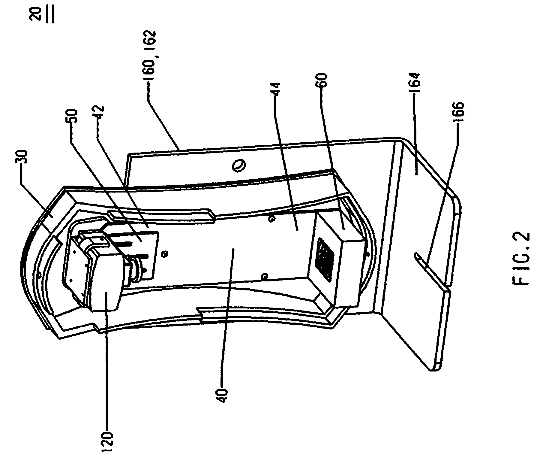
The present invention is a dental X-ray film viewing device that includes a film reader and an image display screen. The film reader has a support panel, a film illumination station, and a video camera. The video camera has a distortion at full field less than 3% and a resolution of at least 512×582. The image display screen is connected to the video camera and displays an enlarged digital image of the X-ray film. The device also includes a system housing for shielding stray light from the environment. The method of producing the enlarged digital image involves placing the X-ray film on a translucent film window, backlighting the film window with a diffused white light source, and using the video camera to capture the image. The enlarged digital image can be visually reviewed and used for dental diagnosis or treatment monitoring. The technical effects of the invention include improved image quality, reduced distortion, and improved dental diagnosis and treatment monitoring.

Problems solved by technology

Furthermore, the objects being radio-graphed, the patients' teeth, are also small.
Consequently, the dental images are exceedingly small, making diagnosis and patient case presentation utilizing these x-ray films a difficult task.
Although the x-ray film has very high resolution, the information revealed often is not sufficient for diagnosis or monitoring a dental treatment because of its small size.
To date, this is an exceedingly expensive technique, and the produced digital images do not have the same image clarity and quality of the x-ray film.
Furthermore, as the digital image produced depends on the algorithm and software used by instrument, reliability of information provided still remains questioned by the dentists and the regulatory authorities.
Up to date, none of these instruments has received U.S. Food and Drug Administration's approval as a diagnostic instrument.
With this device, the back lighting of the x-ray view box directly sheds on to the camera, which causes poor contrast of the digital images produced.
Furthermore, as can be appreciated, with such a shorter working distance, a substantial distortion of the digital image is inherent.
Moreover, this typically produces an image with a focused center portion, but blurred edge portions, therefore, not the entirety of the enlarged image can be used for the purpose of diagnosis.
However, the quality of the digital images produced is poor.
For example, if the x-ray film has an image of five teeth, the teeth at the distal portions where the image has more distortion and poor resolution, can not be used by the dentist.
However, the film tray obstructs the edges of the x-film from displaying.

Method used


Image

  • Dental x-ray film viewing device
  • Dental x-ray film viewing device
  • Dental x-ray film viewing device

Examples


example 1

[0062]The performance of the instant dental x-ray film viewing device is evaluated against a Telerex Video x-ray film viewer using the standard 1″×1.25″ intra-oral x-ray films.

[0063]The instant dental x-ray film viewing device has been described above. In this exemplary embodiment, a micro CCD board camera produced by Samsung, equipped with a Edmundoptics' infinite conjugate MVO®μ-Video™ imaging lens having a focal length of 25.0 mm is used. This micro video lens has a distortion at full field of −0.46%. The working distance is set at about 235 mm, which is substantially longer than that of Telerex. The prior art instrument has a height of 9.4 cm, and estimated working distance of the lens is less than 80 mm. A 15 inch LCD monitor is used as the display screen in the instant device.

[0064]Table 2 shows some of the characteristics of the instant device versus the prior art instrument. More specifically, resolution and diagnostic usefulness of the enlarged image, visibility of the enti...


PUM

PropertyMeasurementUnit
working distanceaaaaaaaaaa
sizeaaaaaaaaaa

Abstract

A dental x-ray film viewing device includes a film reader having a digital video camera and an image display screen connected to the video camera for displaying an enlarged digital image of the x-ray film using signals transmitted directly from the video camera. The film reader includes a film illumination station that has a back-lighted film seat. The film seat includes a translucent plate and a non-transparent film anchoring wafer having an opening complimentary to the x-ray film for seating the x-ray film, and blocking light in an area surrounding the x-ray film. The film reader has a video camera having lens thereof focusing on the film window, and the lens has a distortion at the full field less than 3%. The device provides a substantially enlarged high resolution and high contrast digital image as an accurate representation of the original x-ray film, without digital processing by a computer.

Description

FIELD OF THE INVENTION[0001]The present invention relates to a dental X-ray film viewing device. More specifically, the viewing device provides a substantially enlarged digital image of an entirety of an intra-oral x-ray film using signals transmitted directly from a video camera without digital processing by a computer.BACKGROUND OF THE INVENTION[0002]Intra-oral dental x-ray film is used in general dentistry and by some specialists. By necessity, these films must be small in order to be placed inside of the patients' mouth. Furthermore, the objects being radio-graphed, the patients' teeth, are also small. Consequently, the dental images are exceedingly small, making diagnosis and patient case presentation utilizing these x-ray films a difficult task. After obtaining the x-rays films, the dentist must examine them in order to determine if there are dental diseases to be treated. For example, the dentist identifies dental caries by noting dark (radiolucent) areas on the x-ray image, ...

Claims


Application Information

Patent Timeline
Patent Type & Authority Applications(United States)
IPC IPC(8): A61B6/14G03B42/02G03C5/16A61B6/51
CPCG03B42/08H04N1/00129H04N1/195H04N1/19594A61B6/506H04N2201/0079H04N2201/0084H04N2201/0089H04N2201/0412H04N5/32H04N23/30
Inventor LUCAS, JAMES JOHNMANSSURI, FEREIDON WOLFGANGLUCAS, FRANCIS J.
Owner LUCAS JAMES JOHN
Learn More