Expandable porous mesh bag device and methods of use for reduction, filling, fixation and supporting of bone

a mesh bag and expandable technology, applied in the field of expandable porous mesh bag devices and methods of use for reducing, filling, fixing and supporting bone, can solve the problems of not specifically designed to remove disc tissue, leave behind tissue fragments, and inefficient tools, so as to avoid puncture fear and avoid the effect of slipping

Inactive Publication Date: 2008-04-10
SPINEOLOGY
View PDF92 Cites 124 Cited by
  • Summary
  • Abstract
  • Description
  • Claims
  • Application Information

AI Technical Summary

Benefits of technology

[0010] In cases in which the bone is to be compacted, the methods and devices of this invention employ a catheter tube attached to an inflatable porous fabric bag as described in U.S. Pat. Nos. 5,549,679 and 5,571,189 to Kuslich, the disclosures of which are incorporated herein by reference. Those bags may be inflated with less fear of puncture and leakage of the inflation medium than thin walled rubber balloons. They may also be used over a Scribner balloon to protect the balloon from breakage and eventually seepage.
[0011] The devices of U.S. Pat. Nos. 5,549,679 and 5,571,189 to Kuslich, additionally provide the surgeon with the advantage of safely skipping the first balloon inflation steps of Scribner and Scholten, by expanding the bag through introduction of fill material, such as a bone repair medium thereby correcting the bony defect and deformity and stabilizing it in one step of the procedure.

Problems solved by technology

These tools are very inefficient, as the stringy annular tissues tend to simply move aside and remain attached when these tools are used.
Scalpels and pick-ups tend to leave behind fragments of tissue.
These fragments can lead to re-herniation—a painful condition that might require a second or even a third operation.
Tools, such as those described in the above cited references, while useful, were not specifically designed to remove disc tissue, and tend to require multiple passes to completely clean out the inner annulus tissue.
The use of rongeurs and curettes also tends to leave behind fragments of tissue that may also lead to re-herniation.
Furthermore, because these rongeurs and curettes require multiple passes, the operation may be prolonged, possibly leading to increased bleeding and higher infection rates.
An open section in the middle of the beam helps reduce this movement, but does not effectively eliminate the unwanted movement.
While electrical cautery does effectively destroy disc tissue, it produces heat and smoke in the process.
Heat can injure surrounding tissue, including delicate spinal nerves, potentially causing further harm to the patient.
In addition, the production of smoke may obscure vision and interfere with the surgeons ability to properly perform the operation.
Laser cautery like electrical cautery methods also produce heat and smoke.
Low energy lasers tend to be less effective and therefore the disc removal procedure can be prolonged and less than complete.
Higher energy lasers produce more heat and smoke and therefore can lead to tissue damage beyond the area of intended removal.
But while they are very useful for removing hard tissues, such as bone, they do not efficiently and effectively remove soft tissues, such as disc material.
Rotary burrs attempt to automate and improve the efficiency of disc removal, but these motorized devices are potentially dangerous when used around the spinal cord and spinal nerves as they develop heat, may grab soft tissue and may penetrate too far.

Method used


Image

  • Expandable porous mesh bag device and methods of use for reduction, filling, fixation and supporting of bone
  • Expandable porous mesh bag device and methods of use for reduction, filling, fixation and supporting of bone
  • Expandable porous mesh bag device and methods of use for reduction, filling, fixation and supporting of bone

Examples


Embodiment Construction

[0036] In the following detailed description, similar reference numerals are used to depict like elements in the various figures.

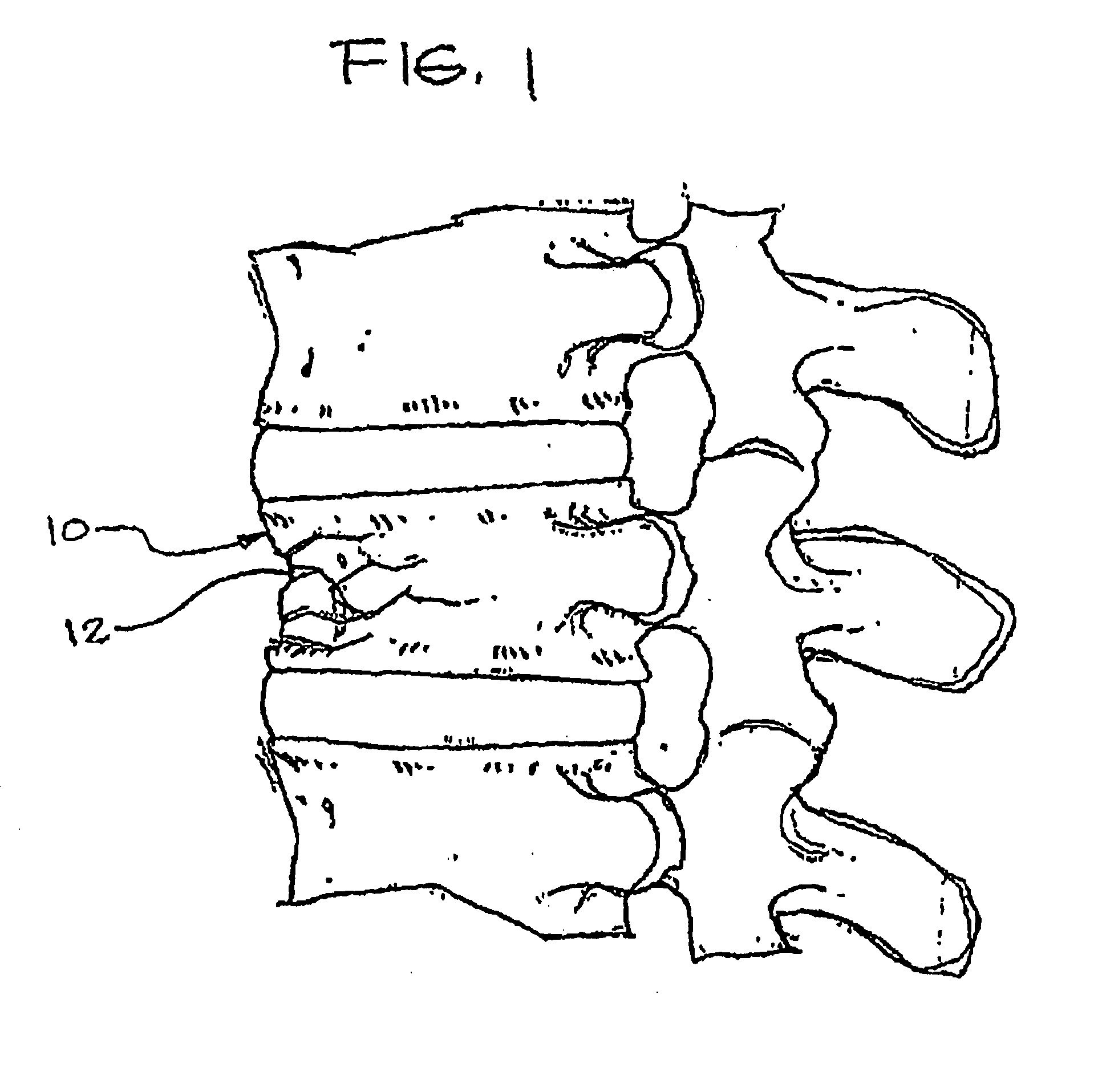
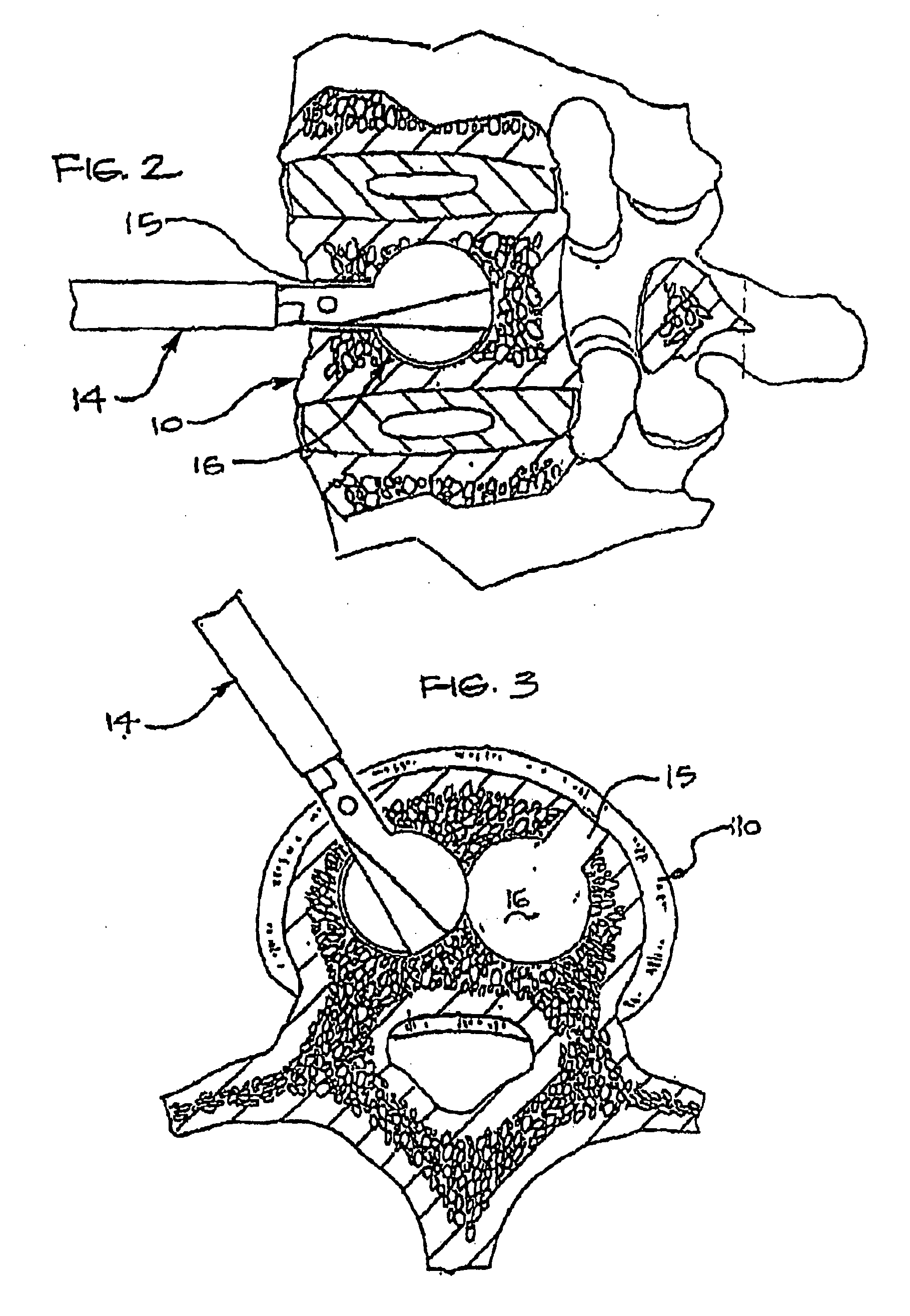
[0037]FIG. 1 shows a typical vertebra 10 having compression fractures 12 that is in need of repair. As indicated above the damaged portion of the vertebra 10 may be reamed out, compacted, or otherwise repaired. For example, FIG. 2 shows a reamer 14 entering the vertebra 10 anteriorly to make an opening 15 and cavity 16. Alternatively, multiple cavities 16 may be formed such as is shown in FIG. 3.

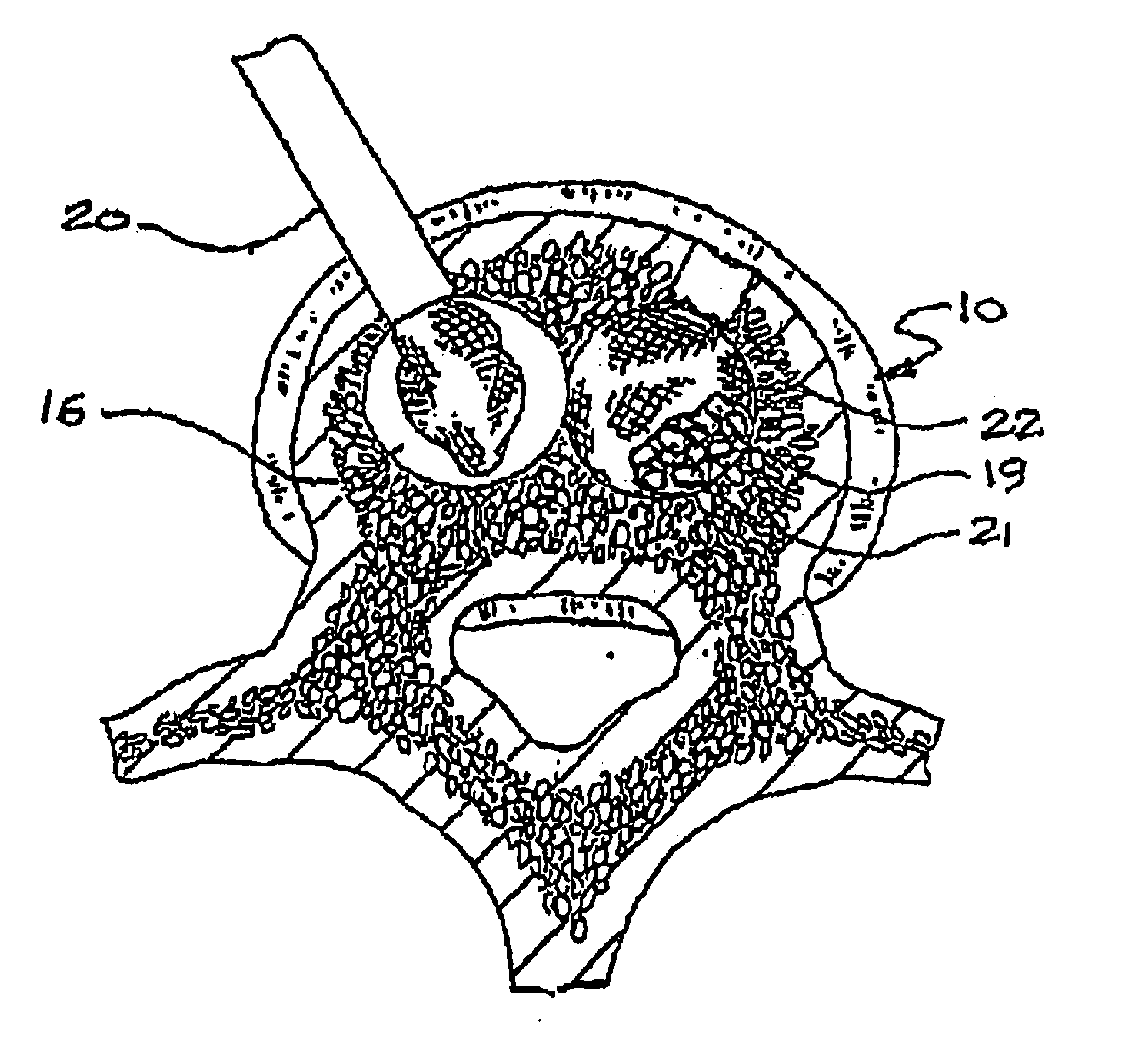
[0038] As previously mentioned, the damaged portion of the vertebra 10 may be compacted in addition to or instead of being reamed out. In FIG. 4, a delivery tube or catheter 20 is seen in the process of delivering an expandable fabric bag 22 into the vertebra 10 or into a cavity 16 present therein. As indicated, the cavity 16 may have been created through reaming, compaction by the bag 22 or other device, or by other means. Once the bag 22 is positioned within th...


PUM


Abstract

A method of treating a compression fracture in a bone comprising the steps of forming a transverse cavity within said bone defined by at least one substantially flat surface lying substantially in a transverse plane formed by and communicating with said transverse cavity, the transverse cavity having a substantially uniform transverse extent and a maximum height, the maximum height being less than said transverse extent and applying a force within said transverse cavity generally normal to said surface to displace said surface and restore said bone to its substantially normal anatomic position.

Description

CROSS-REFERENCE TO RELATED APPLICATIONS [0001] This application is a continuation of Ser. No. 11 / 282,910 filed on Nov. 18, 2005 which is a divisional application of U.S. patent Ser. No. 10 / 440,036, filed May 16, 2003, which claims priority to U.S. patent application Ser. No. 09 / 909,667, filed Jul. 20, 2001, which claims priority to U.S. Provisional Application No. 60 / 219,853 filed Jul. 21, 2000, the entirety of which is incorporated herein by reference.BACKGROUND OF THE INVENTION [0002] This invention relates to methods and devices for correcting bone abnormalities and involves the use of a surgical mesh bag which is inserted into a prepared cavity in bone. The bag is inflated using bone replacement material to expand and fill the cavity. [0003] U.S. Pat. Nos. 5,549,679 and 5,571,189 to Kuslich, describe a device and method for stabilizing the spinal segment with an expandable, porous fabric implant for insertion into the interior of a reamed out disc which is packed with material t...

Claims


Application Information

Patent Timeline
Patent Type & Authority Applications(United States)
IPC IPC(8): A61B17/88A61B17/70A61F2/28A61F2/44
CPCA61B17/00234A61B17/1617A61B17/1628A61B17/1668A61B17/1671A61B17/1675A61B2017/00557A61B17/3472A61B17/7097A61B17/7098A61B17/8855A61B2017/00261A61B17/32002
Inventor KUSLICH, STEPHEN D.KUSLICH, JOHN E.
Owner SPINEOLOGY
Why Eureka
Try Eureka