Methods and Devices for Organ Partitioning

a technology for organs and partitions, applied in medical science, surgical staples, surgery, etc., can solve the problems of difficult placement of staples, t-tags, sutures or other fastening systems from inside the organ or cavity, and the endoscopic partitioning of organs through natural orifices is difficult, so as to achieve the effect of bringing the organ walls together

Inactive Publication Date: 2008-05-29
KELLEHER BRIAN
View PDF45 Cites 57 Cited by
  • Summary
  • Abstract
  • Description
  • Claims
  • Application Information

AI Technical Summary

Benefits of technology

[0009]The preferred methods and devices described herein provide for improved methods and devices for tissue securement, and, in particular, for securing the walls of a hollow organ together. Such securement may be useful for creating partitions within the hollow organ such as for the treatment of obesity involving the stomach. The device may be positioned in the body with the aid of an endoscope and may be used from within the hollow organ to bring the organ walls together easily so that the walls may be secured.

Problems solved by technology

Despite the benefits to the patient, endoscopic partitioning of organs through natural orifices is difficult.
This is primarily because the natural orifices present small volumes where working space is limited; therefore, the placement of staples, t-tags, sutures or other fastening systems is difficult from inside the organ or cavity.
Particularly in the case of gastric stapling, placing a stapler inside the stomach and then securing two opposing walls of the stomach together is difficult when working from only inside the stomach without having access to the inside and the outside walls of the stomach.
It is possible to pull in linear tucks of stomach wall tissue from two opposing walls and to then staple or otherwise affix such segments together, but it has been shown that such couplings are less apt to heal together and therefore hold up over time than couplings that involve stapling the full thickness of the two opposing walls from outside the stomach.

Method used


Image

  • Methods and Devices for Organ Partitioning
  • Methods and Devices for Organ Partitioning
  • Methods and Devices for Organ Partitioning

Examples


Embodiment Construction

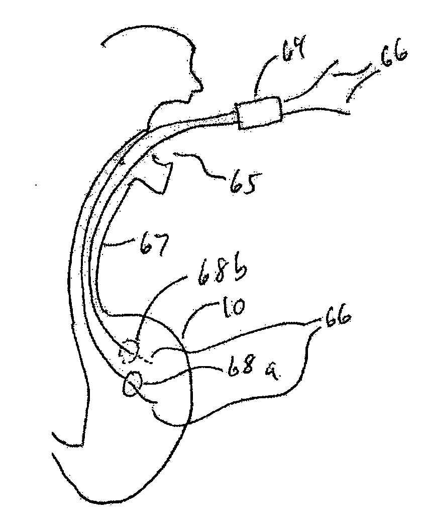
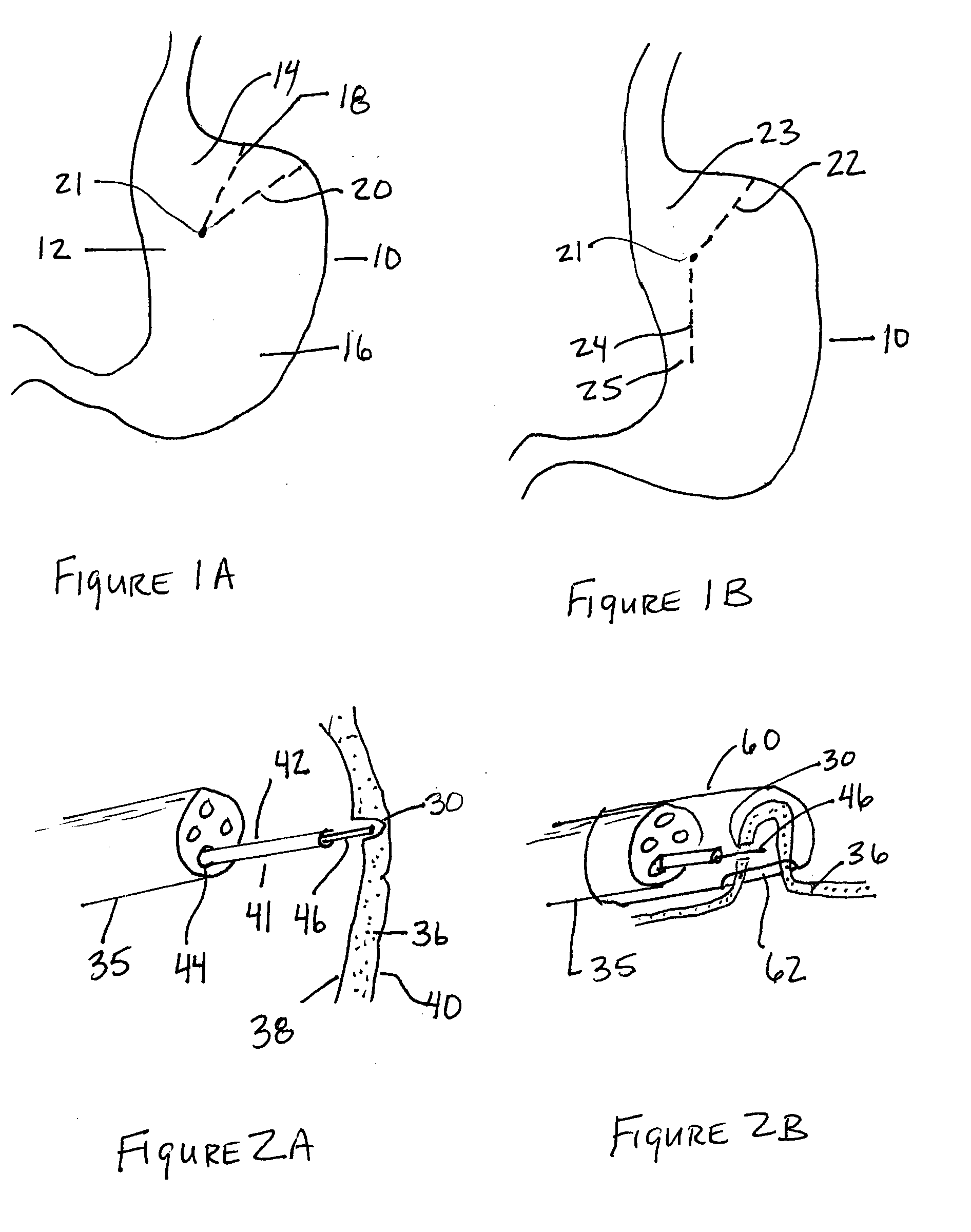
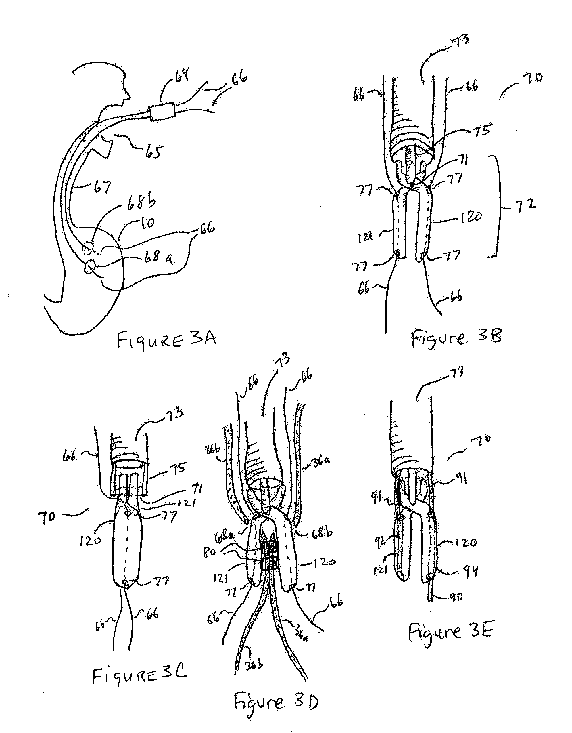
[0038]FIGS. 1-7 depict various embodiments of an organ partitioning device that may be used to secure walls of a hollow organ together in order to create compartments within the hollow organ or to reduce the volume of the organ. The described device may be delivered to the hollow organ with a number of methods including percutaneous, surgical and endoscopic means. Preferably the device is configured for delivery through a natural orifice of the body using flexible endoscopic means.

[0039]The device and method of the present invention may be applicable to many body organs, cavities, lumens or vessels and may be used to treat a wide variety of indications where a secure wall to wall requirement is present. The coupling of one wall of a body lumen to another may be useful in any number of disease states, treatment modalities and body sites. Although this invention and method may be illustrated in this description using the stomach, this is not meant to be limiting in any way. It is anti...


PUM


Abstract

Methods and devices for partitioning hollow organs or cavities of the body are described. A tissue securement device having securement arms is positioned through portals formed in opposing tissue walls of an organ. The securement arms may be articulated to facilitate placement. Once positioned the securement arms may be activated to secure the opposing walls of the organ together. The securement arms may secure the walls together through the use of staples, t-tags, sutures, clamps, helical anchors, braided anchors or fastening devices or systems.

Description

CLAIM OF PRIORITY[0001]This application claims priority under 35 U.S.C. § 119(e) to U.S. Provisional Application No. 60 / 867,332, filed Nov. 27, 2006, the entire contents of which are hereby expressly incorporated by reference.BACKGROUND OF THE INVENTION[0002]1. Field of the Invention[0003]The present invention relates to devices and methods for partitioning a hollow organ of the body. In particular positioning a device through a natural orifice, forming openings in the walls of the organ, positioning a device through these openings and securing the walls of the organ together.[0004]2. Description of the Related Art[0005]Hollow organs or cavities in the body are sometimes secured together in order to create compartments within the hollow organ or to reduce the volume of the organ. These devices are typically delivered to the hollow organ using open surgical or laparoscopic means. For example, the stomach may be partitioned as a treatment for GERD (gastro esophageal reflux disease) or...

Claims


Application Information

Patent Timeline
Patent Type & Authority Applications(United States)
IPC IPC(8): A61B17/11A61B17/34
CPCA61B17/06066A61B17/068A61B17/1114A61B17/29A61B2017/306A61B2017/00278A61B2017/00557A61B2017/22038A61B18/1492
Inventor KELLEHER, BRIAN
Owner KELLEHER BRIAN
Why Eureka
Try Eureka