Method for tissue culture extraction

Active Publication Date: 2008-07-17
EIGEN HEALTH SERVICES LLC
View PDF53 Cites 59 Cited by
  • Summary
  • Abstract
  • Description
  • Claims
  • Application Information

AI Technical Summary

Benefits of technology

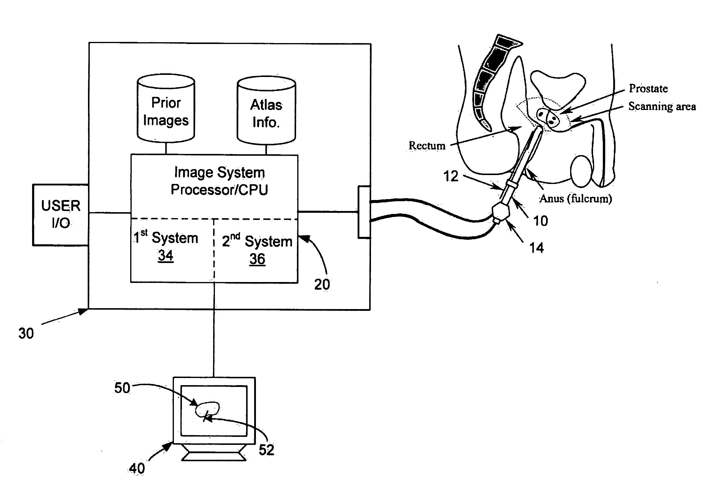
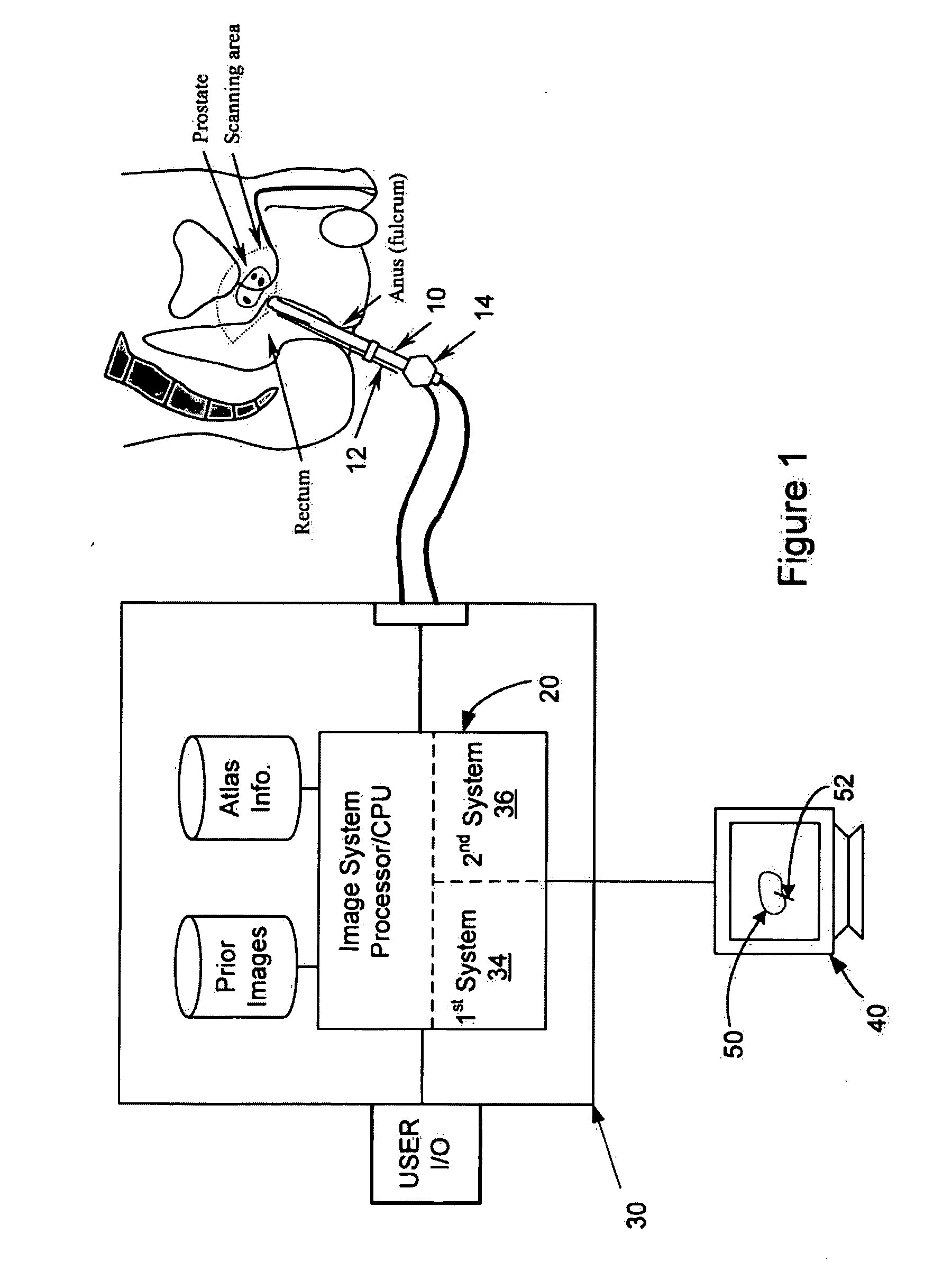
[0005]Provided herein are systems and methods (i.e., utilities) that allow for providing an image guidance system that uses two knowledge-based systems for improving the tissue culture and workflow for urologists. The utility not only uses one knowledge-based system to provide guidelines for biopsy site selection, but also confirms the confidence in site selection through another knowledge-based system. In this regard, the utilities may be implemented in processing systems that are integrated into medical imaging devices and / or that are interconnected to the medical imaging devices and operative to receive data there from. The ability to select and confirm biopsy site selection reduces the need to sample non-suspicions regions and thereby reduces patient discomfort while improving biopsy accuracy. Further such a utility may be performed on-line, for example while a patient remains in view of an imaging device. Accordingly, this may allow for guidance to one or more biopsy sites of interest without repositioning the patient.
[0006]Another relates method is to use a knowledge-based system to guide tissue culture extraction. The acquired data is then fused using multi-modality image data set or warped using a knowledge-based system such as an ATLAS. But a knowledge-based system may only be used as a guideline in the absence of a validation / confirmation system. The present invention presents a new image guidance system for performing biopsy which can overcome the above-referenced problems and improve prostate cancer diagnosis. In presented utility, the suggested biopsy locations from a knowledge-based system are confirmed by another knowledge-based or learning based system such that higher confidence level can be established in selecting the biopsy sites.
[0009]In one arrangement, the second knowledge-based system may perform an image textural analysis and classification for the initial regions of interest. The confirmation procedure may confirm whether the selected target has textural characteristics or other features that are similar to the textural characteristics and or features of histological samples having one or more classified malignancies, tumors and / or cancers. In one arrangement, feature vectors are extracted from the regions of interest by image processing algorithms. The feature vectors may include, without limitation, statistical features, gradient features and / or Gabor filtering features. Features with the most discriminant power may selected through a feature selection algorithm. Further multiple features may be selected for each region of interest. As a result, multiple features from each region may be compared with predetermined features associated with known cancerous, malignant and / or benign histological samples. Where multiple regions of interest are considered, multiple features are extracted and / or multiple features are compared, processing may be performed in parallel processing paths to reduce the processing time required to confirm one or more regions of interest.
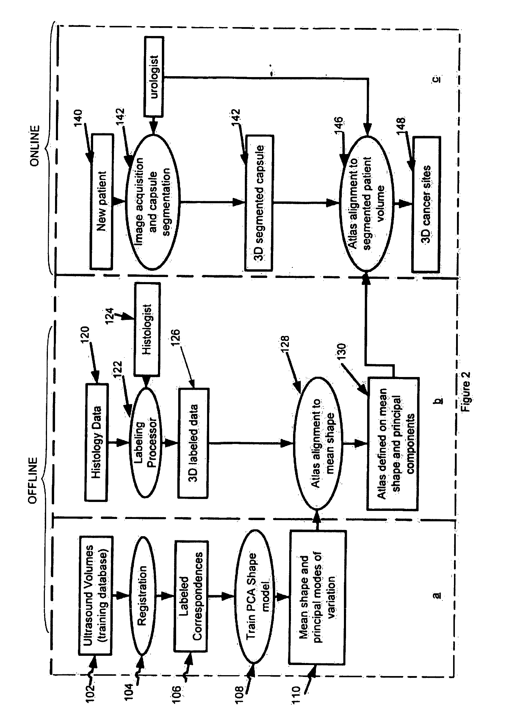
[0010]In accordance with another aspect a system and method (i.e., utility) for training and utilizing a biopsy site confirmation system is provided. The utility may include the following steps, without limitation: (1) generating tumor ground truth locations or regions; (2) extraction of information known to the tumor region (e.g., feature extraction); (3) feature selection; and / or (4) classifier training. The following steps may be included in the online classification procedure of the biopsy site confirmation utility: (1) extracting information known to the suggested biopsy regions (ROIs) which provided by a first knowledge-based system; and / or (2) classification and confirmation of the regions of interest using trained classifier. The classification system may be an online system and can achieve real-time to assist urologists in prostate cancer diagnosis. The multi-threading technique and multi-resolution technique are designed in the workflow.
[0014]In one arrangement, the classifier training procedure can train a system / machine based on the urologists' prior knowledge and known ground truth, so the trained system / machine can be used to classify and confirm a new unknown region in the system. One classification method applied aims at minimizing the bound on the generalization error (i.e., error made by the learning machine on data unseen during training) rather than minimizing the training error over the data set.

Problems solved by technology

However, in TRUS-directed biopsies, tissue sampling within different sections is done in a random fashion, since no prior information about the spatial location of the cancer is available.
This not only causes pain to the patient (55% of men report discomfort during prostate biopsy), but also decreases the accuracy of the method (this method has shown to have a large false negative detection rate ranging from 27% to 39%).

Method used


Image

  • Method for tissue culture extraction
  • Method for tissue culture extraction
  • Method for tissue culture extraction

Examples


Embodiment Construction

[0031]Reference will now be made to the accompanying drawings, which assist in illustrating the various pertinent features of the various novel aspects of the present disclosure. Although the invention is described primarily with respect to an ultrasound imaging embodiment, the invention is applicable to a broad range of imaging modalities and biopsy techniques, including MRI, CT, and PET, which are applicable to organs and / or internal body parts of humans and animals. In this regard, the following description is presented for purposes of illustration and description. Furthermore, the description is not intended to limit the invention to the form disclosed herein. Consequently, variations and modifications commensurate with the following teachings, and skill and knowledge of the relevant art, are within the scope of the present invention.

[0032]With reference to FIG. 1, in the illustrated system the tissue culture / biopsy is assisted by an image guidance system 30. The image guidance ...


PUM


Abstract

An image guidance system is provided for improving tissue culture extraction where the tissue is extracted under the guidance of first and second knowledge-based systems. The first knowledge-based system provides initial suggested regions of interest for tissue biopsy in an internal organ. These regions of interest are then confirmed as being of interest of being benign by the second knowledge-based system. Information from two different sources may provide a more accurate, intelligent and robust method that helps in selecting biopsy sites accurately, such that suspicious regions are not overlooked and the benign regions are not unnecessarily operated upon. This not only helps in better diagnosis and treatment, but also helps reduce pain to the patient, in addition to reducing wastage of resources and invaluable time.

Description

CROSS REFERENCE TO RELATED APPLICATIONS[0001]This application claims priority under 35 U.S.C. §119 to U.S. Provisional Application No. 60 / 884,941 entitled: “An Improved Method For Tissue Culture Extraction” having a filing date of Jan. 15, 2007, the entire contents of which is incorporated by reference herein.FIELD[0002]The present invention relates to medical imaging. One aspect is directed to image guided surgery using a first knowledge-based system to identify regions of interest for biopsy and using a second knowledge-based system to confirm such regions as being desirable for biopsy.BACKGROUND OF THE INVENTION[0003]Prostate cancer is the most common type of cancer found in American men, other than skin cancer. According to American Cancer Society, there will be about 234,460 new cases of prostate cancer in the United States in 2006 and about 27,350 men will die of this disease. Prostate cancer is only curable at an early stage. Therefore, early detection is extremely important ...

Claims


Application Information

Patent Timeline
IPC IPC(8): G06K9/00G06V10/25
CPCA61B8/08A61B8/12G06K9/3233G06K9/6206G06T2207/30081G06T7/0083G06T7/0089G06T2207/10068G06T2207/10136G06T7/0012Y10S128/922G06T7/12G06T7/149G06V10/25G06V10/754
Inventor SURI, JASJIT S.WEI, LIYANGKUMAR, DINESH
Owner EIGEN HEALTH SERVICES LLC
Why Eureka
Try Eureka