

Method for Depositing Radiation in Heart Muscle
a radiation depositing and heart muscle technology, applied in the field of tissue treatment, can solve the problems of limiting the rate at which x-ray images are acquired, blood cannot efficiently empty out of the atria into the ventricles, and radiation exposure of collateral tissues
- Summary
- Abstract
- Description
- Claims
- Application Information
AI Technical Summary
Benefits of technology
Problems solved by technology
Method used
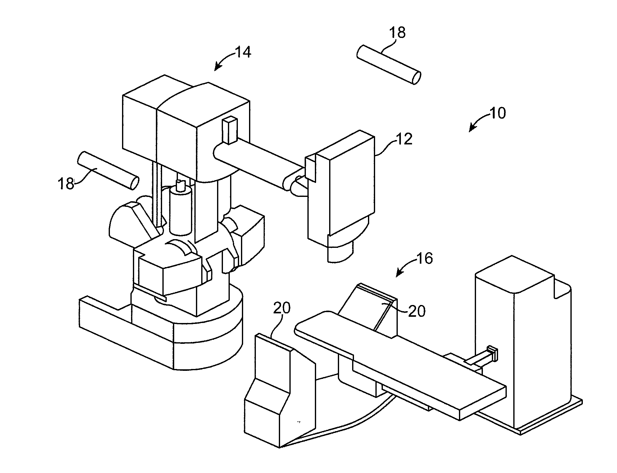
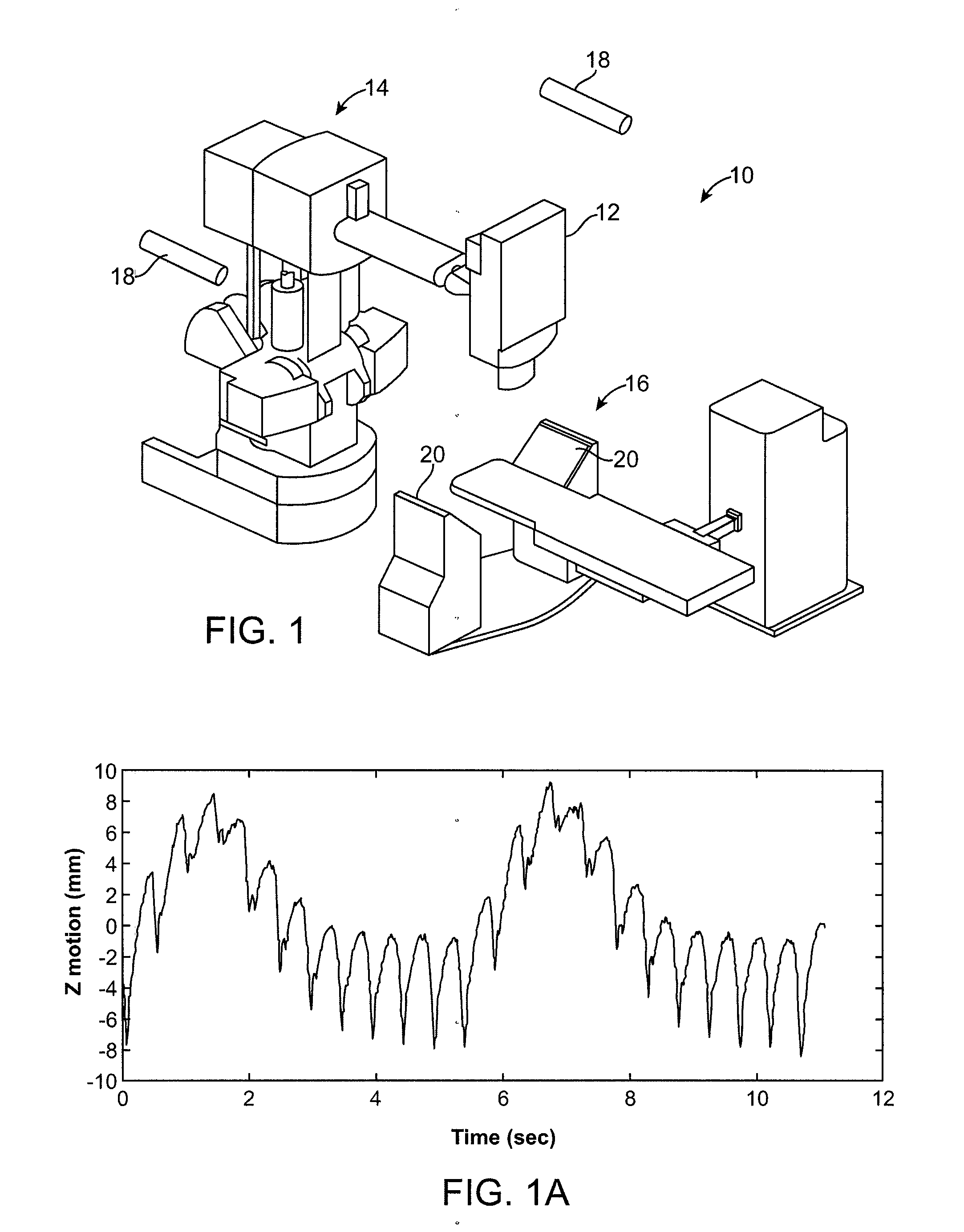
Image

Examples
case 1
[0056] No (or Negligible) Cardiac Component; with Significant Respiratory Component
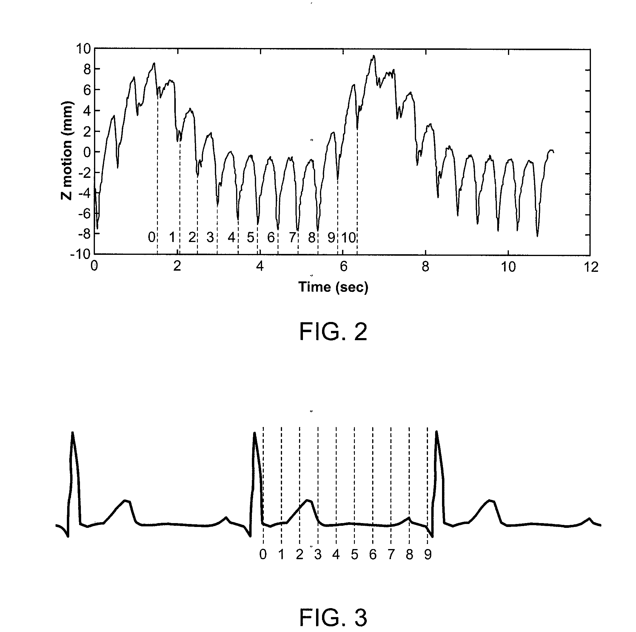
[0057]In this case, the target in the heart muscle has only a respiratory component and not a cardiac component. Targets in the left atrium near the pulmonary veins may fall into this category. The steps may include:[0058]1. Acquire a single CT volume at a cardiac phase, Φ, of the cardiac cycle. Use a high speed CT scanner such as the 64-slice Siemens SOMOTOM Definition to acquire CT volumes quickly, e.g. one volume in 83 ms. Contrast agents may be used. Outline the target in this volume.[0059]2. During patient registration stage, just prior to radiation delivery, acquire a series pairs of N X-rays, X-Rays(i), i=0, . . . , N−1, and N samples of the signals from the LEDs, LEDs(i), over 1 respiratory cycle at the cardiac phase Φ. FIG. 2 shows this scenario with N=11.[0060]3. For each i=0, . . . , N−1, register X-Rays(i) with the CT volume by correlating DRRs with X-rays(I) using a similarity measure or ...
case 2
[0075] With Significant Cardiac Component and with Significant Respiratory Component
[0076]The target in the heart muscle has both a respiratory component and a cardiac component. Targets in the ventricles near the valves fall into this category.
Approach 1:
[0077]1. Acquire a series of M CT volumes, CT(j), j=0, . . . , M−1, of the heart over one cardiac cycle with the patient holding his / her breath. Use a high speed CT scanner such as 64-slice Siemens SOMOTOM Definition to acquire CT volumes quickly, e.g. one volume in 83 ms. Contrast agents may be used.
2. FIG. 3 shows a typical EKG waveform with M=10 phases where 10 CT volumes are acquired. Outline the target in each of these M volumes. Alternatively, outline the target in one CT volume and automatically track it over all the CT volumes to generate the targets in other CT volumes.
3. Pick one of the CT phases, Φ, as the reference phase. Acquire a series of pairs of N X-rays, X-rays(i), i=0, . . . , N−1, and N samples of the signals fr...
PUM
 Abstract
Description
Claims
Application Information