Method for implanting a cardiovascular valve

Active Publication Date: 2008-07-24
THE CLEVELAND CLINIC FOUND
View PDF44 Cites 373 Cited by
  • Summary
  • Abstract
  • Description
  • Claims
  • Application Information

AI Technical Summary

Benefits of technology

[0006]In one aspect of the present invention, a method is provided for implanting a valve having at least one valve leaflet within the cardiovascular system of a subject. One step of the method comprises preparing a substantially dehydrated bioprosthetic valve and providing an expandable support member having oppositely disposed first and second ends and a main body portion extending between the ends. The substantially dehydrated bioprosthetic valve is attached to the expandable support member so that the substantially dehydrated bioprosthetic valve is operably secured within the main body portion of the expandable support member. The expandable support member is then crimped into a compressed configuration and placed at a desired location within the cardiovascular system of the subject.
[0007]In another aspect of the present invention, a method is provided for implanting a valve having at least one valve leaflet within the cardiovascular system of a subject. One step of the method comprises preparing a substantially dehydrated bioprosthetic valve and providing an expandable support member having oppositely disposed first and second ends and a main body portion extending between the ends. The substantially dehydrated bioprosthetic valve is attached to the expandable suppo

Problems solved by technology

The use of chemically-treated implantable bioprosthetic devices presents several drawbacks, however.
For example, the presence of chemically cross-linked/preserved agents, such as glutaraldehyde, presents an environmental hazard to the operating room personnel who are exposed to these chemicals as well

Method used


Image

  • Method for implanting a cardiovascular valve
  • Method for implanting a cardiovascular valve
  • Method for implanting a cardiovascular valve

Examples


Embodiment Construction

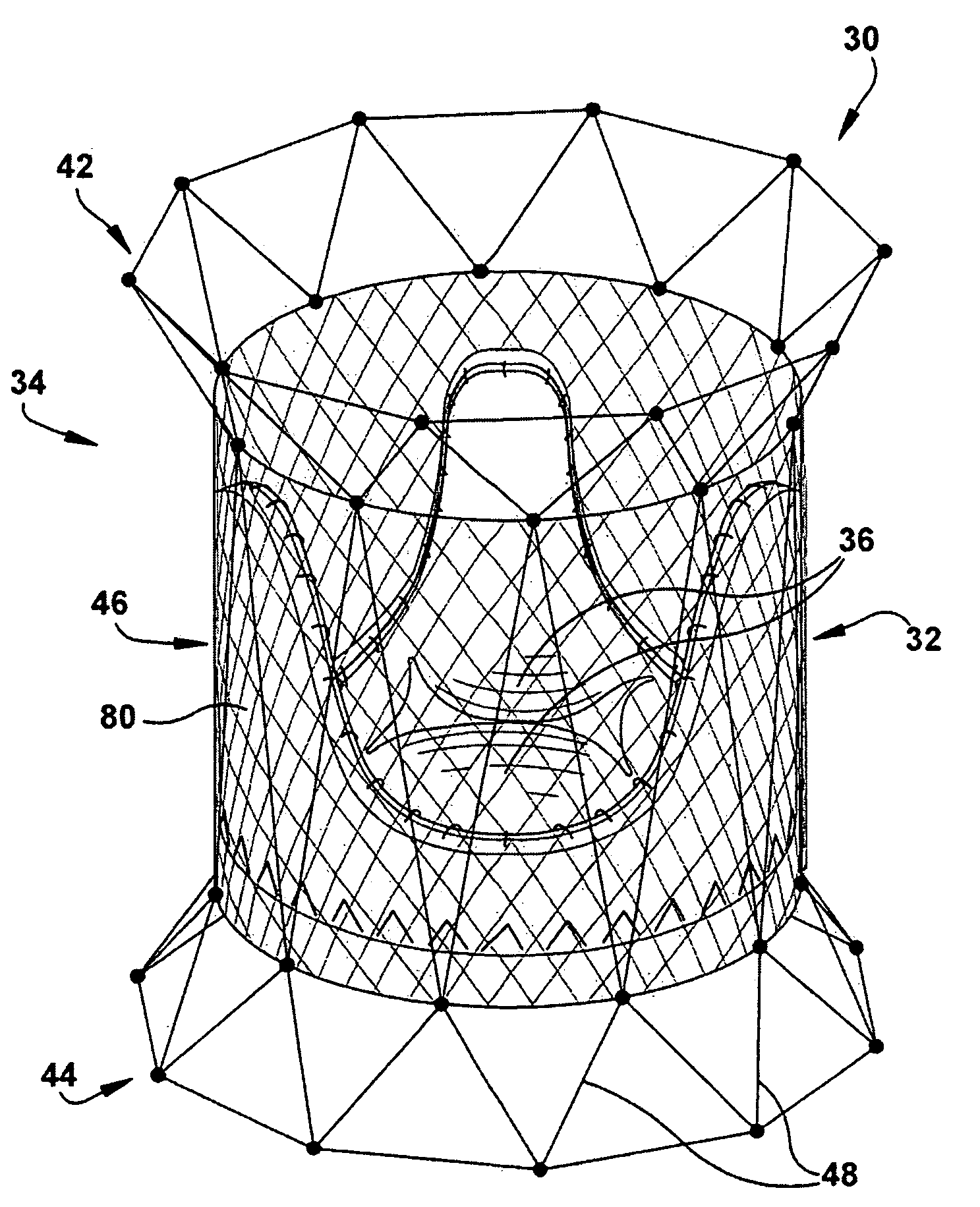
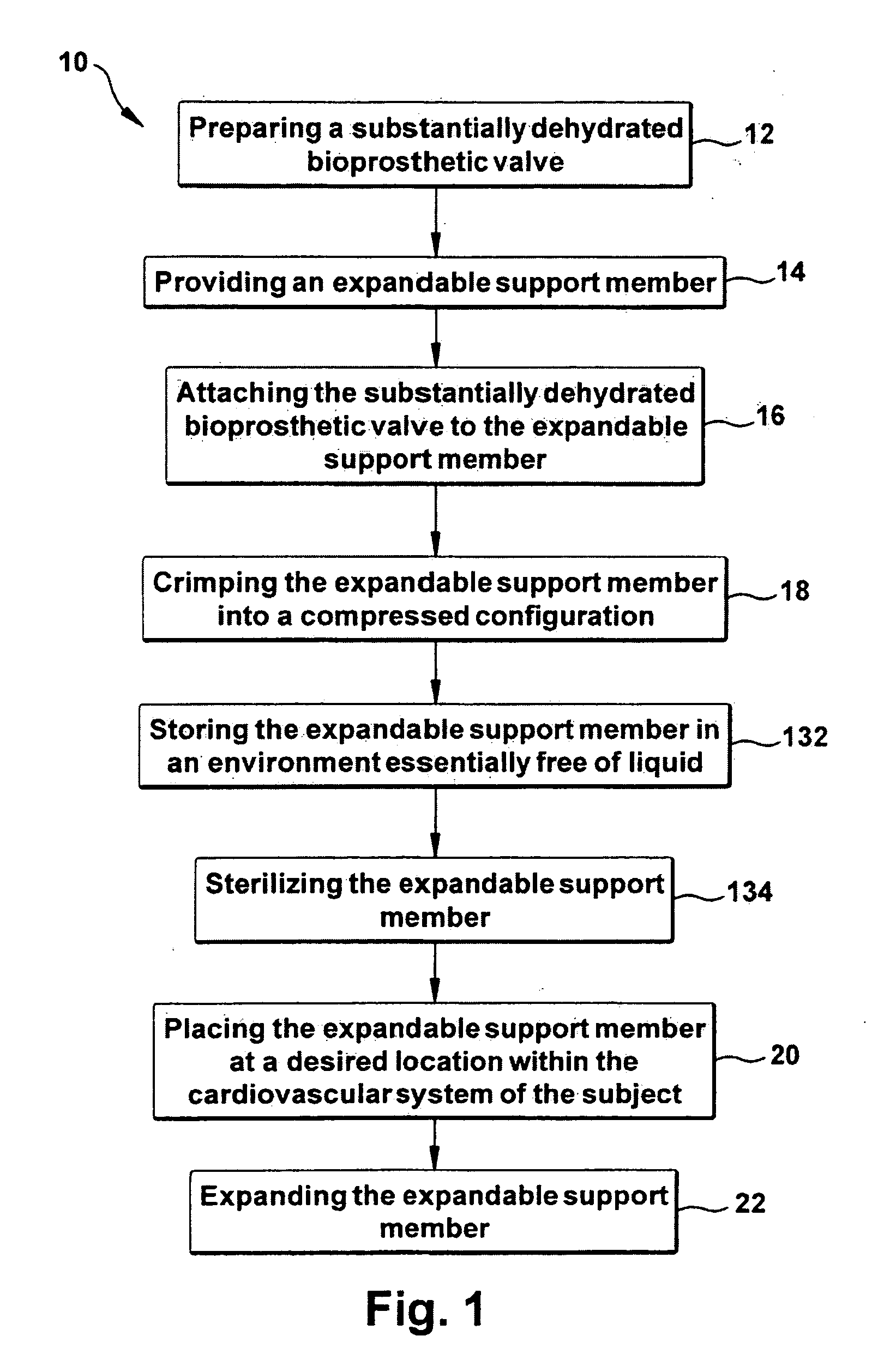
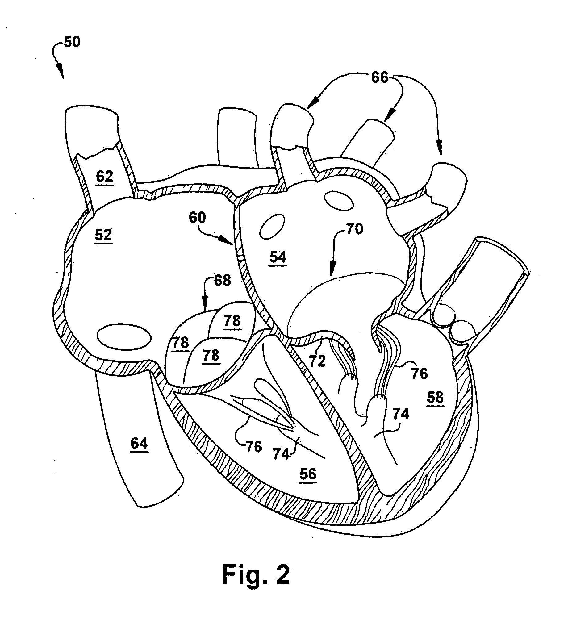
[0038]The present invention relates generally to a method for treating and improving the function of cardiovascular valves, and more particularly to a method for implanting a cardiovascular valve within the cardiovascular system of a subject. As used herein, the term “cardiovascular system” refers to a bodily system consisting of the heart, blood vessels, and blood that circulates blood throughout the body, delivers nutrients and other essential materials to cells, and removes waste products. As representative of the present invention, FIG. 1 illustrates a method 10 for implanting a valve 30 (FIG. 3) having at least one valve leaflet 36 within the cardiovascular system of a subject, wherein the valve comprises a substantially dehydrated bioprosthetic valve 32 securely attached to an expandable support member 34. Advantageously, the method 10 (FIG. 1) of the present invention minimizes, if not eliminates, the possibility of introducing aldehydes into the blood stream of the subject w...


PUM


Abstract

A method is provided for implanting a valve having at least one valve leaflet within the cardiovascular system of a subject. One step of the method includes preparing a substantially dehydrated bioprosthetic valve and then providing an expandable support member having oppositely disposed first and second ends and a main body portion extending between the ends. Next, the substantially dehydrated bioprosthetic valve is attached to the expandable support member so that the substantially dehydrated bioprosthetic valve is operably secured within the main body portion of the expandable support member. The expandable support member is then crimped into a compressed configuration and placed at a desired location within the cardiovascular system of the subject. Either before or after placement at the desired location, fluid or blood re-hydrates the substantially dehydrated bioprosthetic valve.

Description

RELATED APPLICATION [0001]This application claims priority from U.S. provisional patent application Ser. No. 60 / 881,244, filed on Jan. 19, 2007, the subject matter of which is incorporated herein by reference.TECHNICAL FIELD [0002]The present invention relates generally to a method for treating and improving the function of cardiovascular valves, and more particularly to a method for implanting a cardiovascular valve within the cardiovascular system of a subject.BACKGROUND OF THE INVENTION [0003]A number of implantable bioprosthetic devices are currently being used for treating patients with cardiovascular diseases and defects. Such implantable devices are useful for replacing diseased, damaged, or congenitally malformed components of a patient's cardiovascular system. Thus, damaged or diseased heart valves have been replaced with chemically-fixed, bioprosthetic heart valves prepared from tissues of porcine or bovine origin. Similarly, regions of damaged or diseased blood vessels ma...

Claims


Application Information

Patent Timeline
IPC IPC(8): A61F2/24A61F2/04
CPCA61F2/2418A61F2/2433A61F2/2436A61F2002/9522Y10T29/49925A61F2/243A61F2220/0008A61F2230/0078A61F2250/0092A61F2/9522A61F2/9524A61F2/2415A61F2/0095A61L27/3625A61L27/3629A61L27/3687A61L27/3691
Inventor NAVIA, JOSE LUISCHEN, JI-FENG
Owner THE CLEVELAND CLINIC FOUND
Why Eureka
Try Eureka