Method of Compounding and Ultrasound Image

a technology of ultrasound image and compounding method, which is applied in the field of compounding ultrasound image, can solve problems such as deteriorating the quality of ultrasound image, and achieve the effects of smooth ultrasound image, clearer tissue contour view, and reduced speckle noise level

Inactive Publication Date: 2008-08-14
SAMSUNG MEDISON
View PDF5 Cites 58 Cited by
  • Summary
  • Abstract
  • Description
  • Claims
  • Application Information

AI Technical Summary

Benefits of technology

[0010]Since the present invention combines image frames obtained with several different transmission frequencies to provide a compound ultrasound image, it can reduce a speckle noise level and form a smoother ultrasound image.
[0011]Further, the ultrasound image, which is compounded in accordance with the present invention, can provide a clearer view of tissue contours and can present even narrow vessels and muscular tissues.
[0012]Furthermore, as frequency compounding is used together with spatial compounding, a synergistic effect is provided. Therefore, a high-resolution image can be obtained.

Problems solved by technology

However, the ultrasound image acquired through such conventional method generally contains speckles, which considerably deteriorate the quality of the ultrasound image.

Method used


Image

  • Method of Compounding and Ultrasound Image
  • Method of Compounding and Ultrasound Image
  • Method of Compounding and Ultrasound Image

Examples


Embodiment Construction

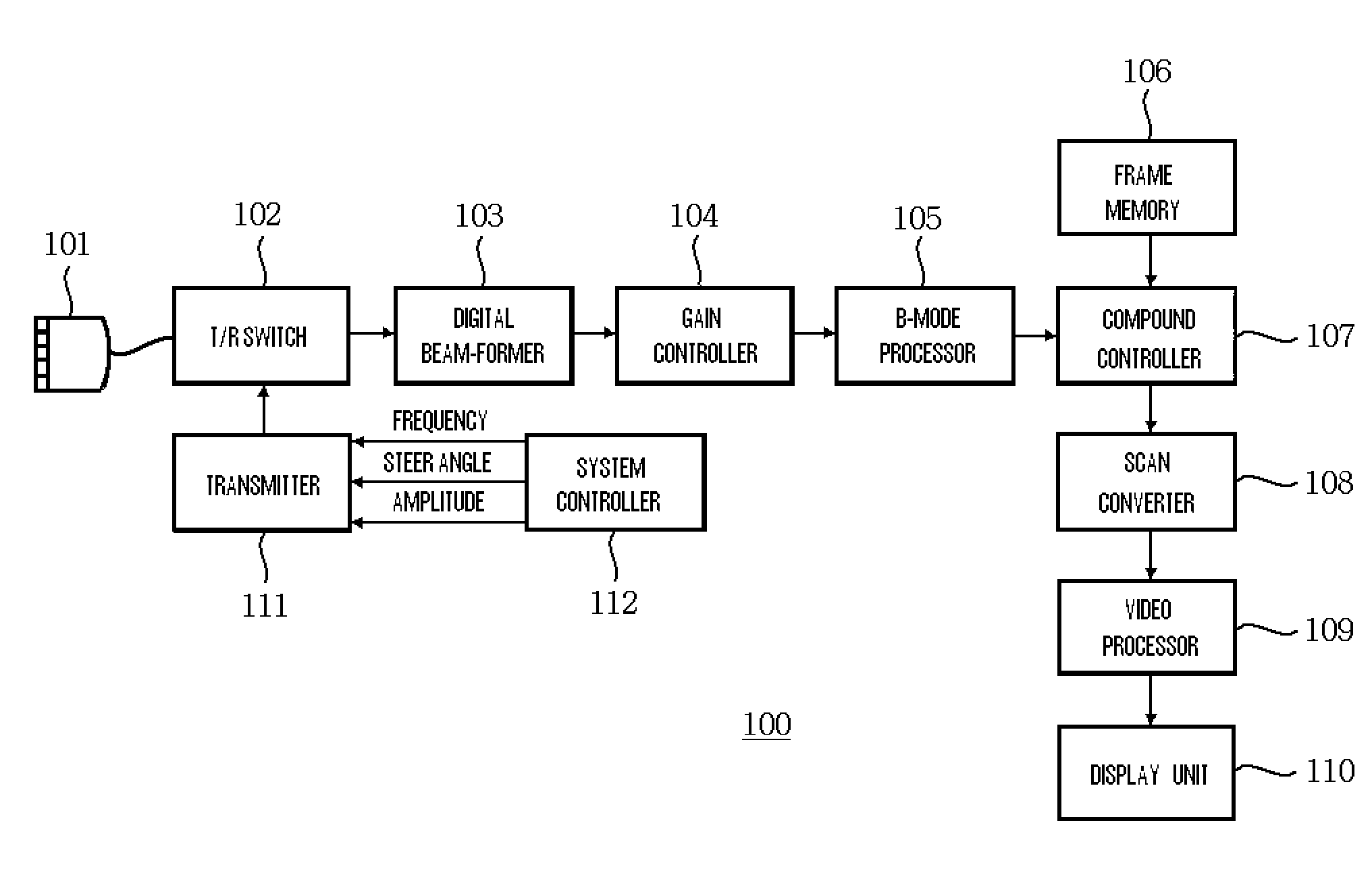
[0017]FIG. 1 is a functional block diagram of an illustrative ultrasound image display apparatus constructed in accordance with an embodiment of the present invention.

[0018]Referring to FIG. 1, the ultrasound image display apparatus 100 includes: a scan header 101 having a transducer array; a transmit / receive (T / R) switch 102; a transmitter 111; a system controller 112; a digital beam-former 103; a gain controller 104; a brightness-mode (B-mode) processor 105; a frame memory 106; a compound controller 107; a scan converter 108; a video processor 109; and a display unit 110.

[0019]The system controller 112 determines the frequency and amplitude of ultrasound signals and a steer angle at which the ultrasound signals are to be transmitted. The transmitter 111 generates ultrasound signals based on the information determined by the system controller 112. The scan header 101 with the transducer array is responsible for transmission of the generated ultrasound signals and reception of signa...


PUM


Abstract

In a method of compounding an ultrasound image, ultrasound signals having a transmission frequency are transmitted to a target object at a predetermined steer angle. Then, signals reflected by the target object are received. Based on the received signals, an image frame is formed. By repeating the above steps with different transmission frequencies, two or more image frames are obtained. The obtained image frames are then combined to provide a compound ultrasound image. The steer angle varies based on the transmission frequency.

Description

TECHNICAL FIELD[0001]The present invention generally relates to a method of compounding an ultrasound image, and more particularly to a method of compounding an ultrasound image through the use of spatial compounding and frequency compounding having a plurality of different transmission frequencies.BACKGROUND ART[0002]Ultrasound imaging systems are widely used in the medical diagnostic field since they can obtain an image of a target object through non-invasive means, i.e., by transmitting ultrasound signals to the object and processing their reflection. Conventional three-dimensional (3D) ultrasound imaging systems have an array of ultrasound transducers or probes, which can generate ultrasound pulses and receive echo signals of the ultrasound pulses reflected off an object. In a conventional method, the transducers transmit ultrasound signals of a fixed frequency and then receive signals reflected by a target object. In such a method, only one frequency component corresponding to ...

Claims


Application Information

Patent Timeline
Patent Type & Authority Applications(United States)
IPC IPC(8): A61B8/00
CPCG01S7/52046A61B8/5207G01S15/8995G01S7/52092A61B8/00
Inventor LEE, KWANG JUKIM, JONG SIKKIM, CHEOL AN
Owner SAMSUNG MEDISON
Why Eureka
Try Eureka