
Image Processing Method for a Microscope System

a microscope system and image processing technology, applied in the field of image processing methods, can solve the problems of early death, difficult prenatal diagnosis, labor-intensive analysis of fluorophore-labeled samples, etc., and achieve the effect of significant subjectivity

Inactive Publication Date: 2008-09-04
IKONISYS INC
View PDF6 Cites 22 Cited by
  • Summary
  • Abstract
  • Description
  • Claims
  • Application Information

AI Technical Summary

Benefits of technology

[0018]Embodiments are disclosed which perform various image processing functions which may be employed to implement the automated fluorescence in situ hybridization method. The embodiments include an auto-exposure method for acceptably imaging all regions of the sample over an intensity range exceeding the dynamic range of the digital electronies; a method for fluorescence in situ hybridization (FISH) object of interest enumeration which loc

Problems solved by technology

In contrast, an abnormal number of autosomes typically results in early death.
Although Down syndrome is easily diagnosed after birth, prenatal diagnosis is problematic.
Despite the above-described advances in the development of fluorescent in situ hybridization methods for the diagnosis of genetic abnormalities, the analysis of the fluorophore-labeled sample remains labor-intensive and involves a significant level of subjectivity.
As will be readily apparent, the above-described procedure is quite time-consuming.
Moreover, because the results are visually-perceived, the frequency of erroneous results can vary from one experiment to the next, as well as from one observer to the next.

Method used


Image

  • Image Processing Method for a Microscope System
  • Image Processing Method for a Microscope System
  • Image Processing Method for a Microscope System

Examples


Embodiment Construction

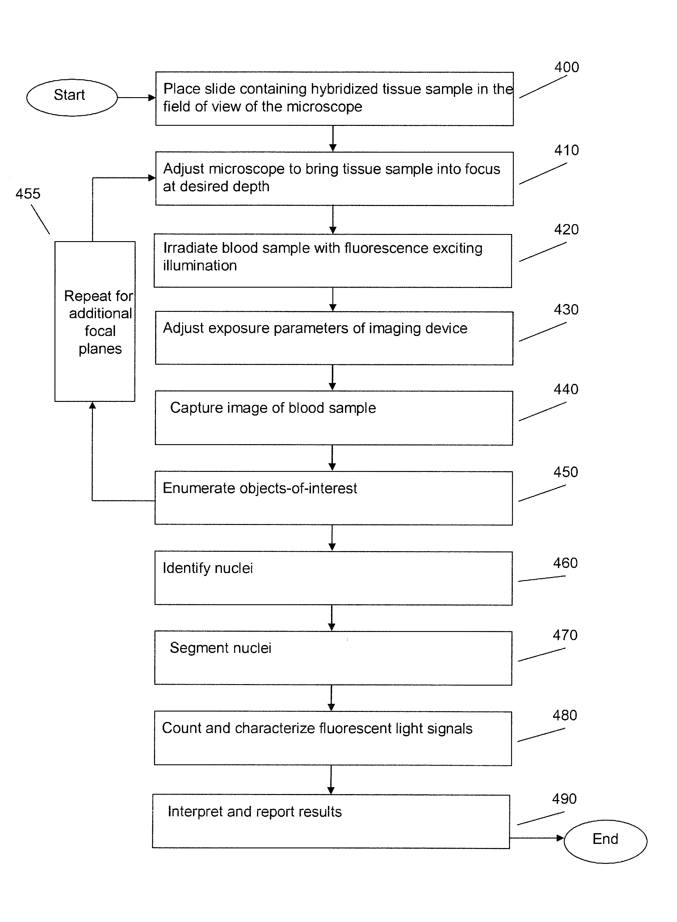
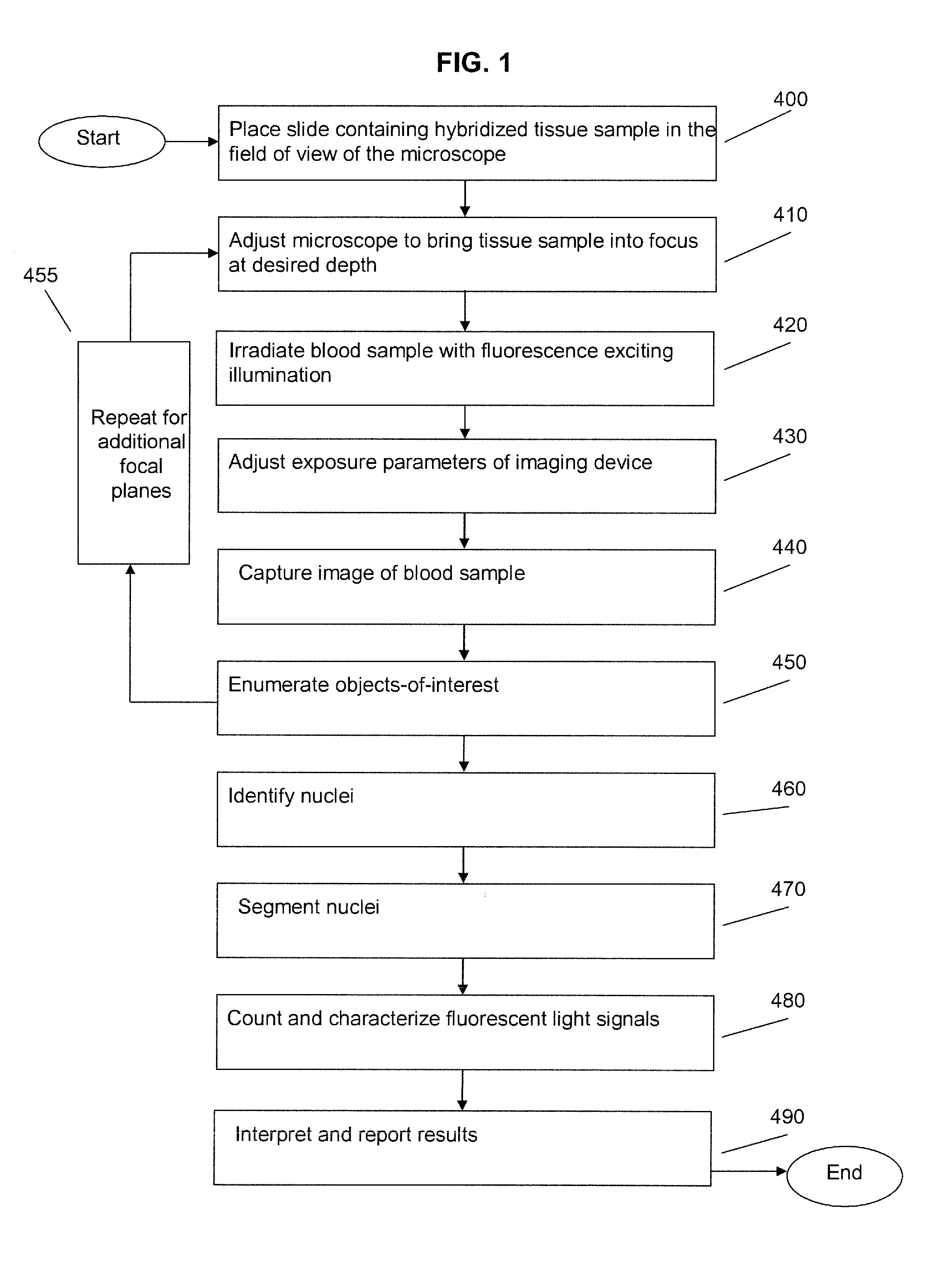
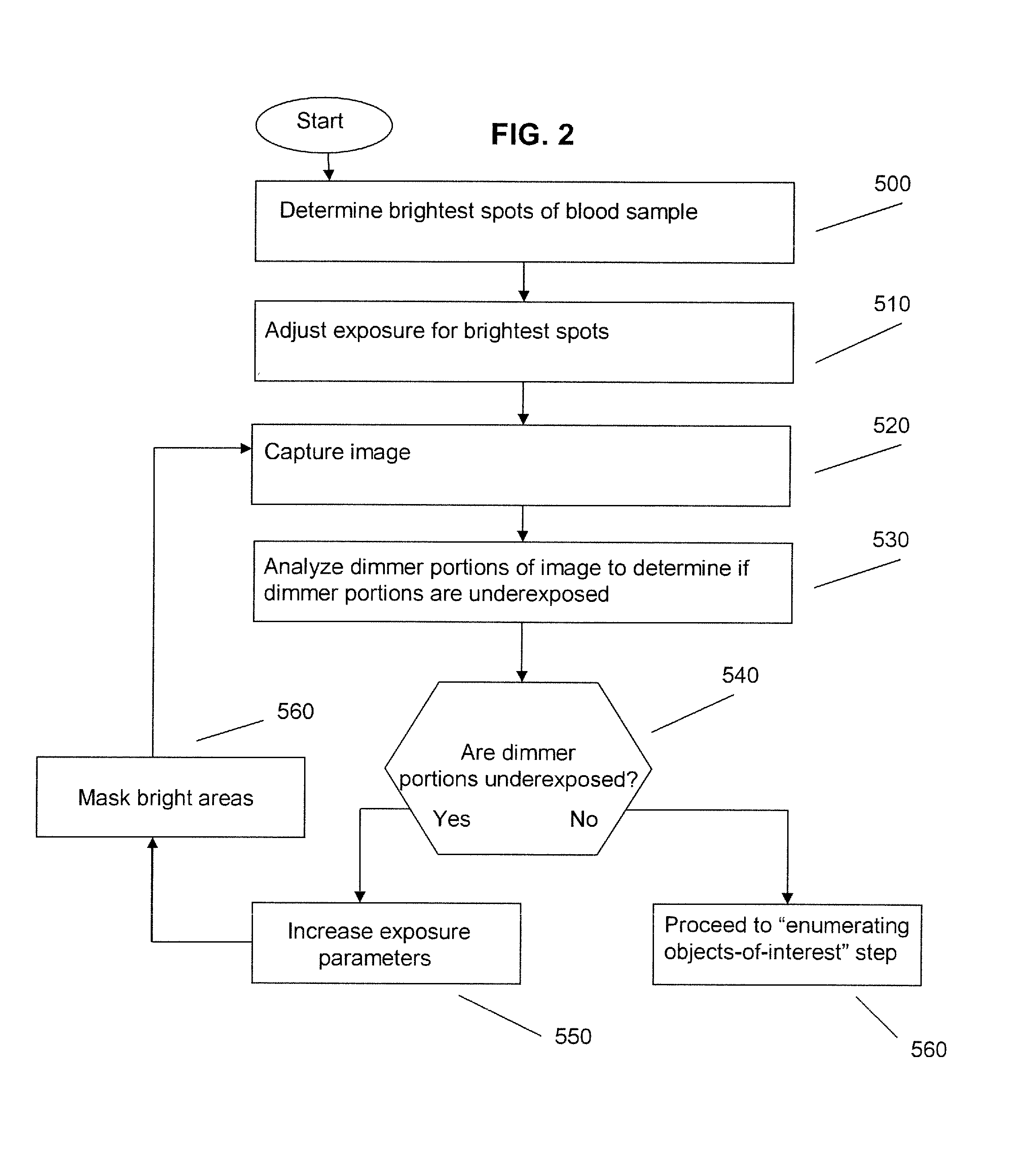
[0034]Auto-Exposure: The acquisition of the digital image typically requires proper exposure of all of the regions of the specimen being examined. The electronic imaging device may be a multi-pixel planar array of light-sensitive detectors in a charge coupled device (CCD), or complementary metal oxide semiconductor (CMOS) elements, or any other technology suitable for converting an optical image into electrical signals. Exemplary CCD cameras include intensified CCD cameras utilizing gating techniques to achieve gate speeds of less than about nine nanoseconds with improved quantum efficiency (e.g., such as Princeton Instruments (Trenton, N.J.) PIMAXMG which support a full range of 16-bit scientific-grade CCDs), allowing for a very fast response limited by the time constant of the output phosphor, and electron-bombarded CCD (EBCCD) wherein photons are detected by a photocathode and released electrons are accelerated across a gap and impact on the hack side of a CCD (allowing for addit...


PUM


Abstract

An embodiment is disclosed for performing the image processing for analyzing the results of a fluorescence in situ hybridization (FISH) microscopic automated sample analysis to determine specific chromosomal characteristics.

Description

[0001]This application claims priority from U.S. Provisional Application Ser. No. 60 / 821,536 filed Aug. 4, 2006. All references cited in this specification, and their references, are incorporated by reference herein where appropriate for teachings of additional or alternative details, features, and / or technical background.BACKGROUND OF THE INVENTION[0002]1. Field of the Invention[0003]The present invention generally relates to image processing methods employed for performing automated microscopic analysis for fluorescence in situ hybridization (FISH) detection of genetic characteristics.[0004]2. Description of the Related Art[0005]Conventional optical microscopy generally employs a microscope slide to which the normal human complement of chromosomes consists of the sex chromosomes (designated X and Y) and 22 autosomes (numbered 1-22). It has been estimated that a minimum of 1 in 10 human conceptions has a chromosome abnormality. As a general rule, an abnormal number of sex chromosom...

Claims


Application Information

Patent Timeline
IPC IPC(8): G06K9/00
CPCG01N21/6458G01N33/48
Inventor ZHU, YANNINGTAFAS, TRIANTAFYLLOSKIM, YOUNGMINWANG, XIUZHONG
Owner IKONISYS INC
Learn More