Clinical utilization of contrast agents to define specific areas within the myocardial wall to provide guidance and localization for ablation, cyroablation, or other techniques in patients with post myocardial infarction

a technology of contrast agents and myocardial wall, which is applied in the field of clinical utilization of contrast agents to define specific areas within the myocardial wall, and can solve the problems of high risk of sudden cardiac death in patients with impaired left ventricular function, high risk of ventricular arrhythmia, and patients with coronary artery disease with prior myocardial infarction. , the cost of implantation of an icd is relatively high

Inactive Publication Date: 2008-09-18
GENERAL ELECTRIC CO
View PDF3 Cites 11 Cited by
  • Summary
  • Abstract
  • Description
  • Claims
  • Application Information

AI Technical Summary

Problems solved by technology

Medical patients with impaired left ventricular function are at high risk of sudden death from ventricular arrhythmias.
Medical patients with coronary artery disease with prior myocardial infarction may experience sudden cardiac death during the first year due to ventricular tachycardia or fibrillation.
The implantation of an ICD, however, is relatively expensive.
There are large numbers of patients with impaired ventricular function, and to implant ICDs in all would place a considerable burden on the health care system.
In addition, the procedure also may expose the patient and the physician to relatively high doses of x-ray.

Method used


Image

  • Clinical utilization of contrast agents to define specific areas within the myocardial wall to provide guidance and localization for ablation, cyroablation, or other techniques in patients with post myocardial infarction
  • Clinical utilization of contrast agents to define specific areas within the myocardial wall to provide guidance and localization for ablation, cyroablation, or other techniques in patients with post myocardial infarction
  • Clinical utilization of contrast agents to define specific areas within the myocardial wall to provide guidance and localization for ablation, cyroablation, or other techniques in patients with post myocardial infarction

Examples


Embodiment Construction

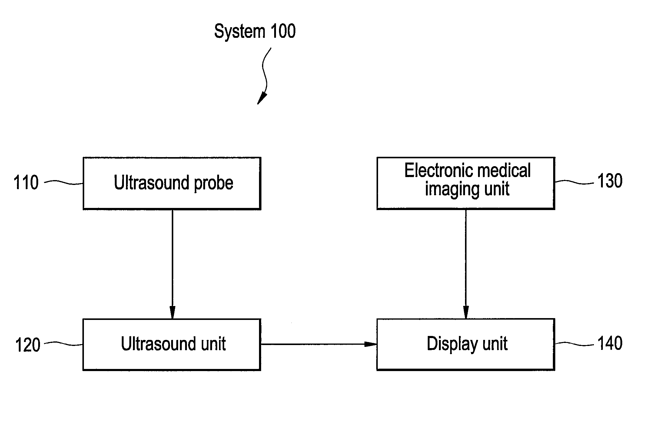
[0019]FIG. 1 illustrates a system 100 for locating non-viable cardiac tissue. The system 100 includes an ultrasound probe 110. The ultrasound probe 110 may be equipment or software that receives data and ultrasound images, including data regarding non-viable cardiac tissue. The system 100 also includes an ultrasound unit 120. The ultrasound unit 120 may be equipment or software that processes data and ultrasound images obtained from the ultrasound probe 110. The system 100 also includes an electronic medical imaging unit 130. The electronic medical imaging unit 130 may represent any equipment or software that permits electronic medical images, such as x-rays, ultrasound, CT, MRI, gated MRI, EBT, MR, or nuclear medicine for example, to be electronically acquired, stored, or transmitted for viewing and operation. The electronic medical imaging unit 130 may receive input from a user. The electronic medical imaging unit 130 may be used to acquire electronic medical images of non-viable ...


PUM


Abstract

Certain embodiments of the present invention provide systems and methods for ablating non-viable cardiac tissue. In an embodiment, the method may include locating non-viable cardiac tissue utilizing an electronic medical image. The method may also include locating non-viable cardiac tissue utilizing an ultrasound unit, wherein a contrast agent is used to characterize the non-viable cardiac tissue. The method may also include guiding a catheter to the location of the non-viable cardiac tissue according to the imaging of the ultrasound unit. The method may also include ablating the non-viable cardiac tissue. The ablating of the non-viable cardiac tissue may include cauterizing the non-viable cardiac tissue, cyroablating the non-viable cardiac tissue, or include radiofrequency ablation. The method may also include attempting to locate the non-viable cardiac tissue utilizing an electronic medical image after ablating the non-viable cardiac tissue.

Description

RELATED APPLICATIONS[0001][Not Applicable]FEDERALLY SPONSORED RESEARCH OR DEVELOPMENT[0002][Not Applicable]MICROFICHE / COPYRIGHT REFERENCE[0003][Not Applicable]BACKGROUND OF THE INVENTION[0004]The present invention generally relates to a method for using contrast agents to define specific areas within the myocardial wall. Specifically, the present invention relates using contrast agents to define specific areas within the myocardial wall to provide guidance for ablation, cyroablation, or other techniques or procedures.[0005]Medical patients with impaired left ventricular function are at high risk of sudden death from ventricular arrhythmias. Non-congenital ventricular arrhythmias are most often caused by an area of living myocardial tissue almost completely surrounded by non-viable cardiac tissue. Cardiac tissue has the unique property of being able to self-stimulate. Tissue areas surrounded by non-viable cardiac tissue do not receive the stimulus from the surrounding tissues. Theref...

Claims


Application Information

Patent Timeline
Patent Type & Authority Applications(United States)
IPC IPC(8): A61B8/00A61B18/18
CPCA61B8/481A61B2018/0262A61B18/1492
Inventor DONALDSON, BRENDA L.CHODAKAUSKAS, THADDEUS H.VADODARIA, SACHIN
Owner GENERAL ELECTRIC CO
Why Eureka
Try Eureka