Method And Apparatus For Blood Vessel Parameter Determinations

a blood vessel and parameter determination technology, applied in the field of blood flow diagnosis, can solve the problems of affecting the measurement flow, affecting the measurement accuracy, and requiring more equipment, so as to achieve the effect of improving the accuracy of blood flow measurement, and reducing the cost of mr method

Inactive Publication Date: 2008-10-23
PAIEON INC
View PDF3 Cites 127 Cited by
  • Summary
  • Abstract
  • Description
  • Claims
  • Application Information

AI Technical Summary

Benefits of technology

[0008]It is an object of the present invention to provide a novel method and apparatus for the determination of coronary reserve and relative coronary reserve of a specific coronary artery segment and other flow related measurements. In accordance with the present invention, there is thus provided a method for determining the arterial reserve of a subject having a blood flow of a velocity below or equal to a maximal velocity value, the method comprising the following steps: receiving one or more first models, each model representing one or more substantially fixed segments of one or more arteries of the subject at a plurality of points in time associated with and during one or more parts of one or more first heart beat cycles, the artery segment having a proximal cross section and a distal cross section; injecting contrast agent into the artery of the subject, when the subject is in a non-hyperemic state, the contrast agent is injected at an injection area having a proximal cross section and having a distance from the substantially fixed segment; determining one or more first parameters from one or more first angiograms representing the substantially fixed artery segment, said first angiogram taken form a projection angle; injecting the subject with substance that simulates hyperemia; receiving one or more second models, representing the substantially fixed segment of the artery of the subject at a plurality of points in time associated with and during one or more parts of one or mode second heart beat cycles; injecting the contrast agent to the artery of the subject, said subject being in a hyperemic state, said contrast agent is injected at the injection area; determining one or more second parameters from one or more second angiograms representing the substantially fixed segment, said second angiogram taken form a projection angle; and determining the arterial reserve as the ratio between one of the first parameters or a combination thereof and one of the second parameters or a combination thereof. The first parameter can be a ratio between the maximal velocity value of blood within the artery segment when the subject is at a hyperemic state and the distance between the substantially fixed segment and the injection area. The second parameter can be a ratio between the maximal velocity value of blood within the artery segment when the subject is at a non-hyperemic state and the distance of the artery segment from the injection area. Within the method, determination of the first parameters can comprise the steps of: determining from the first angiogram taken at a predetermined projection angle and the first model, a density curve for the artery segment; obtaining a velocity profile of the blood flow within the artery; performing curve fitting for the density curve to determine the first parameter. Within the method, determination of the second parameter can comprise the steps of: determining from the second angiogram taken at a predetermined projection angle and second model, a density curve for the at least one artery segment; obtaining a velocity profile of the blood flow within the artery; performing curve fitting for the density curve to determine the second parameter. The distance between the injection area and the substantially fixed segment can be the distance between the distal cross section of the injection area and a cross section of the substantially fixed artery segment located at equal distances from the proximal cross section and from the distal cross section of the substantially fixed artery segment. The method can further comprise the step of creating the first or the second models of the artery of the subject. The first or the second models can be three-dimensional models. The method can further comprise the step of determining the projection angle and the volumes of the fixed segment of the artery of the subject at a plurality of points in time associated with and during the one or more parts of the first heart beat cycle, from the first model and the step of determining the projection angle and the volumes of the fixed segment of the at least one artery of the subject at a plurality of points in time associated with and during the one or more parts of the second heart beat cycle, from the second model. The method can further comprise the step of compensating for the non-perpendicularity of the substantially fixed segment of the artery of the subject. The method can further comprise the step of registering the first angiogram with the first model or the second angiogram with the second model. The method can further comprise a step of determining TIMI grades from local gray level curves in multiple points of the artery. The method can further comprise the step of determining a relative arterial reserve as the ratio between the arterial reserve determined for a first artery segment and the arterial reserve determined for a second artery segment. The first artery segment can be diseased or suspect as being diseased and the second artery segment can be healthy. The arterial reserve can be arterial coronary reserve. The contrast agent injection can performed during the systole of the subject, or continuously throughout an integer number of heart beat cycles of the subject. The contrast agent can be injected radially.
[0009]Another aspect of the present invention relates to a method for determining the blood flow output of a subject having a blood flow having velocity values below or equal to a maximal velocity, the method comprising the steps of: receiving one or more models, representing one or more substantially fixed segments of one or more arteries of the subject at a plurality of points in time associated with and during one or more parts of one or more heart beat cycles; injecting contrast agent into the artery of the subject at an injection area having a distance from the substantially fixed segment; determining from one or more angiograms taken at a projection angle and the model, a density curve for the substantially fixed segment; obtaining a velocity profile of the blood flow within the substantially fixed segment; performing curve fitting for the density curve to determine one or more parameters; substituting the at parameters in the velocity profile to determine velocity values; and integrating the velocity values over a cross section of the substantially fixed segment to obtain the arterial segment output values of the blood flow within the substantially fixed segment of the artery. Within the method, the parameter can be the ratio between the maximal velocity of blood within the substantially fixed segment and the distance of the sub

Problems solved by technology

Extra intervention procedures are required, which makes the Doppler catheter or the pressure wire methods more costly, more invasive, and therefore more risky.
In addition, the extra wire or catheter affect the flow itself and therefore impair the measured flow.
Yet another drawback of the Doppler catheter and the pressure wire is that the catheter or wire must be accurately aligned, otherwise the results are impaired.
The MR method is also costly and physicians are reluctant to use it.
A limitation of the DSA methods is the difficulty in calibrating the relation between the gray level and the volume.
There are too many parameters and not enough available information to accurately

Method used


Image

  • Method And Apparatus For Blood Vessel Parameter Determinations
  • Method And Apparatus For Blood Vessel Parameter Determinations
  • Method And Apparatus For Blood Vessel Parameter Determinations

Examples


Embodiment Construction

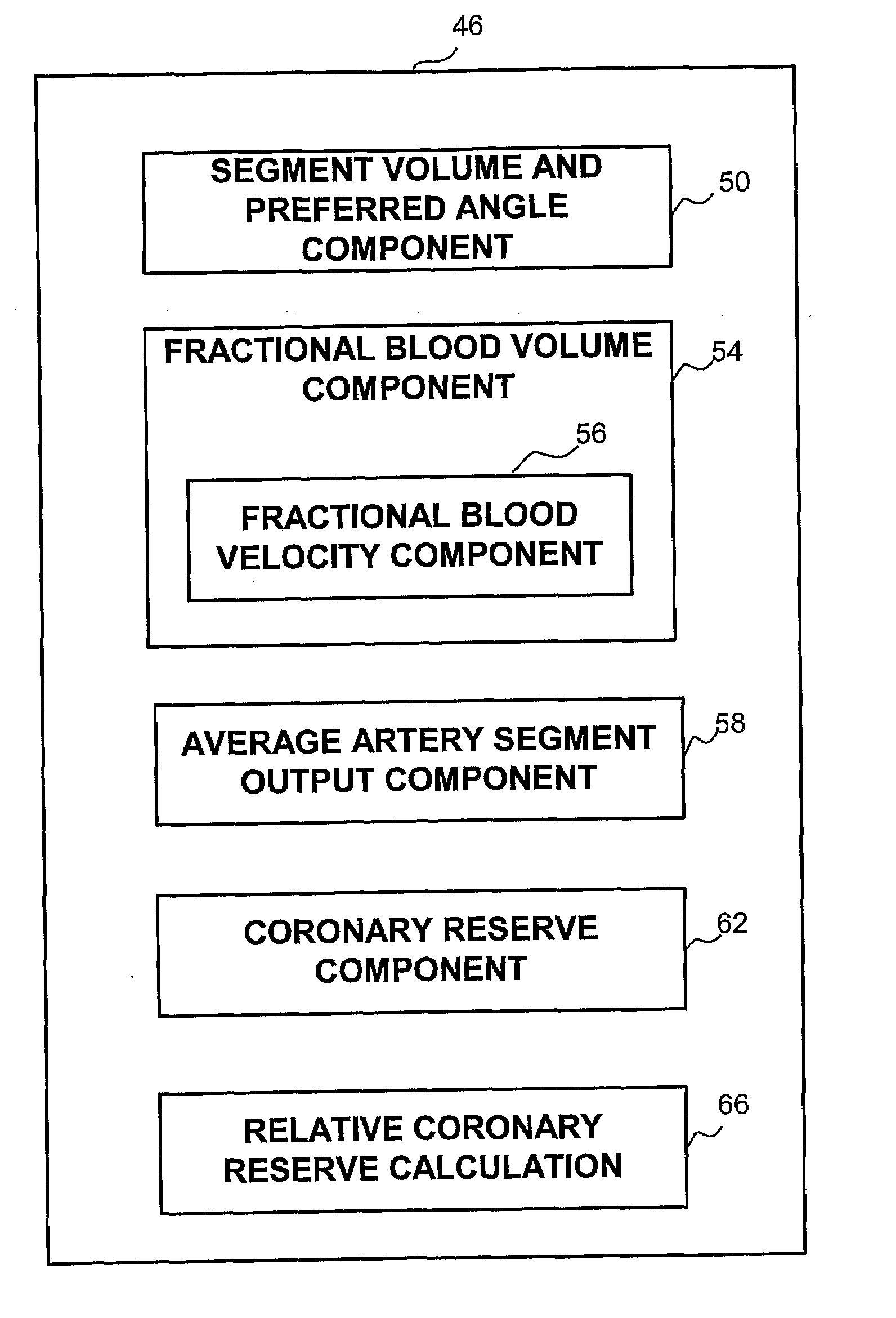
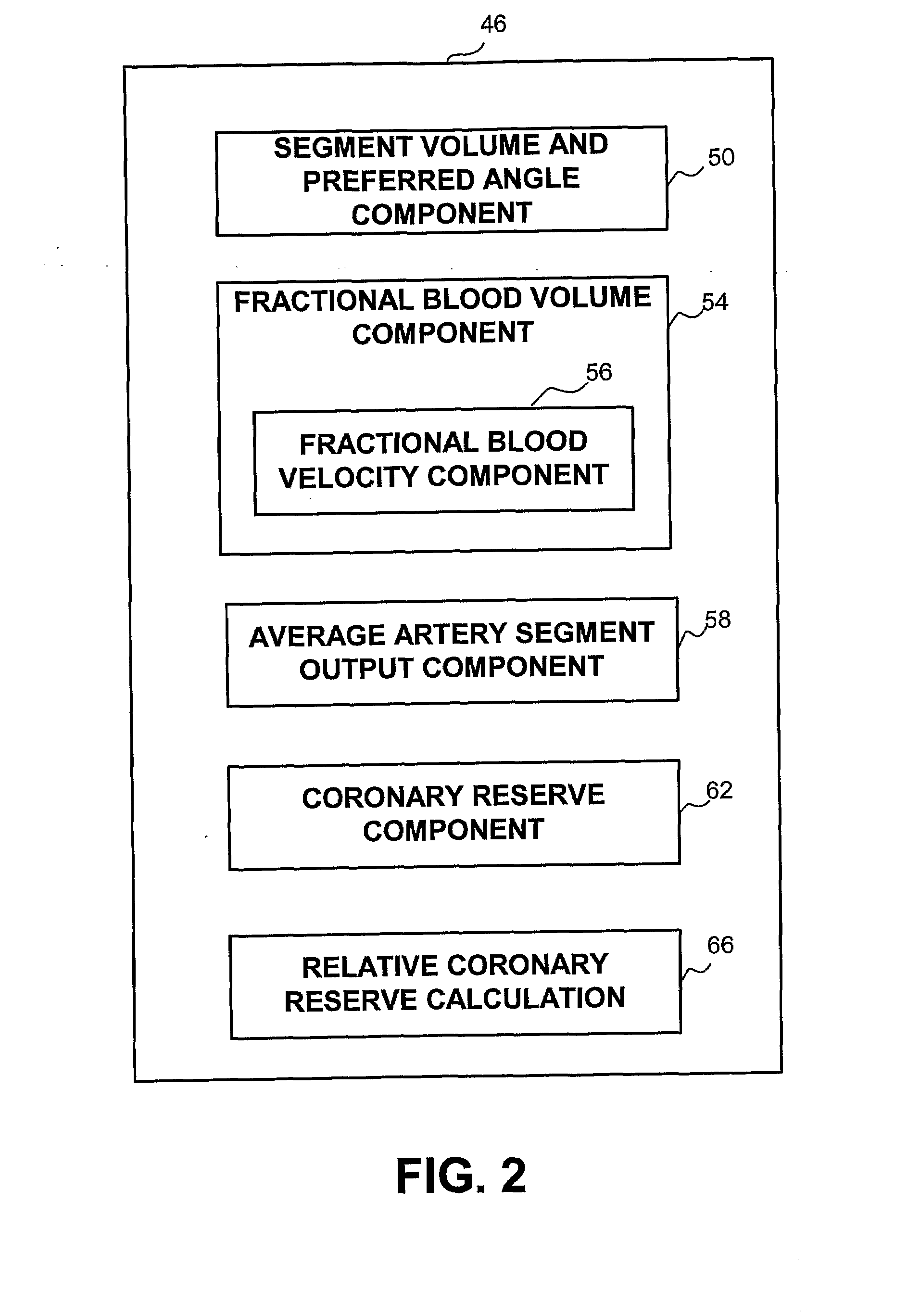
[0029]A new and novel apparatus and method for the determination of coronary reserve and relative coronary reserve and other flow related measurements of a specific coronary artery is disclosed. The proposed method and apparatus first determines the 3D model, including the volume of a fixed segment of an artery at multiple points of time throughout a heart beat cycle. Alternatively, the method uses extrapolation of the 3D model, including the associated volumes from one time segment to another. In yet another alternative, the 3D model and is volumes are received from another source, such as CardiOp by Paieon of Rosh Ha'ayin, Israel. Then, using a contrast agent injected to the subject, the apparatus measures the rate at which the injected material fills the segment of the artery examined. The filling rate and the volume of the segment of the artery examined at the same point in time relatively to the heart beat cycle are used to determine the velocity of the blood flow at that point...


PUM


Abstract

An apparatus and method for the determination of the flow, the coronary reserve and relative coronary reserve of a specific coronary artery. The apparatus and method employ a three-dimensional model (50) providing the volume of a segment of the artery at a plurality of points in time, and disclose various options (56) for determining the arterial flow through the artery segment during one or more heart beat cycles or parts thereof. The determination of the coronary reserve (62) and relative coronary reserve (64) follow from the volume (54) and the flow through the artery. Alternatively, the coronary reserve is determined directly from a velocity profile relating to one or more fixed artery segments and the three-dimensional model.

Description

BACKGROUND OF THE INVENTION[0001]1. Field of the Invention[0002]The present invention relates to blood flow diagnosis in general, and to a method and system for determining the coronary reserve and the relative coronary reserve, in particular.[0003]2. Discussion of the Related Art[0004]The coronary velocity reserve (CVR) also known as the coronary flow reserve (CFR), and generally referenced hereinafter as coronary reserve, is defined to be the ratio between the maximal and the resting coronary blood flow. The maximal coronary blood velocity occurs at times of highly intensive exercise, or it is induced when the subject is injected with coronary vasodilator such as adenosine. The coronary reserve represents the ability of the coronary arteries to supply the excess blood needed to comply with the excessive pumping requirements, relatively to normal (resting) conditions. The normal coronary reserve is greater than 3.0 and in some individuals is greater than 5.0. When the CFR is impair...

Claims


Application Information

Patent Timeline
IPC IPC(8): A61B5/0275A61B6/00
CPCA61B6/507A61B5/0275
Inventor ASSIS, TSURIELKUPER, ASHERZARKH, MICHAELKLAIMAN, MOSHE
Owner PAIEON INC
Why Eureka
Try Eureka